Cemental Tear
This patient was referred to me for examination and consultation regarding a mandibular anterior tooth that had been previously treated by the Referring Dentist. The 49 year old gentleman had a history of trauma when young and discoloration of the crown. The tooth was otherwise virgin.
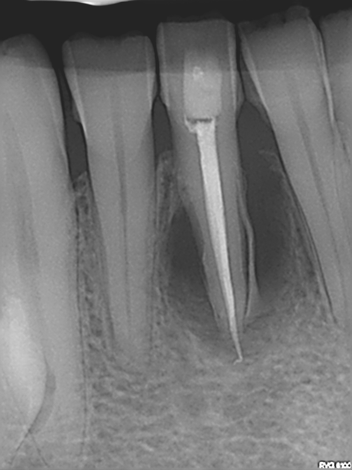
Although the Endodontic treatment appeared to be relatively good (aside from the overextension of the filling material) , a large radiolucent finding (encompassing the entire root in all directions and up the mesial side) had resulted in high mobility and a persistent mucosal defect at the buccal apex.
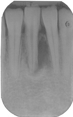
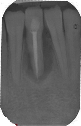
Figures 1 and 2 : Referrial’s Images
Pre and postop images. Treatment by the referral.
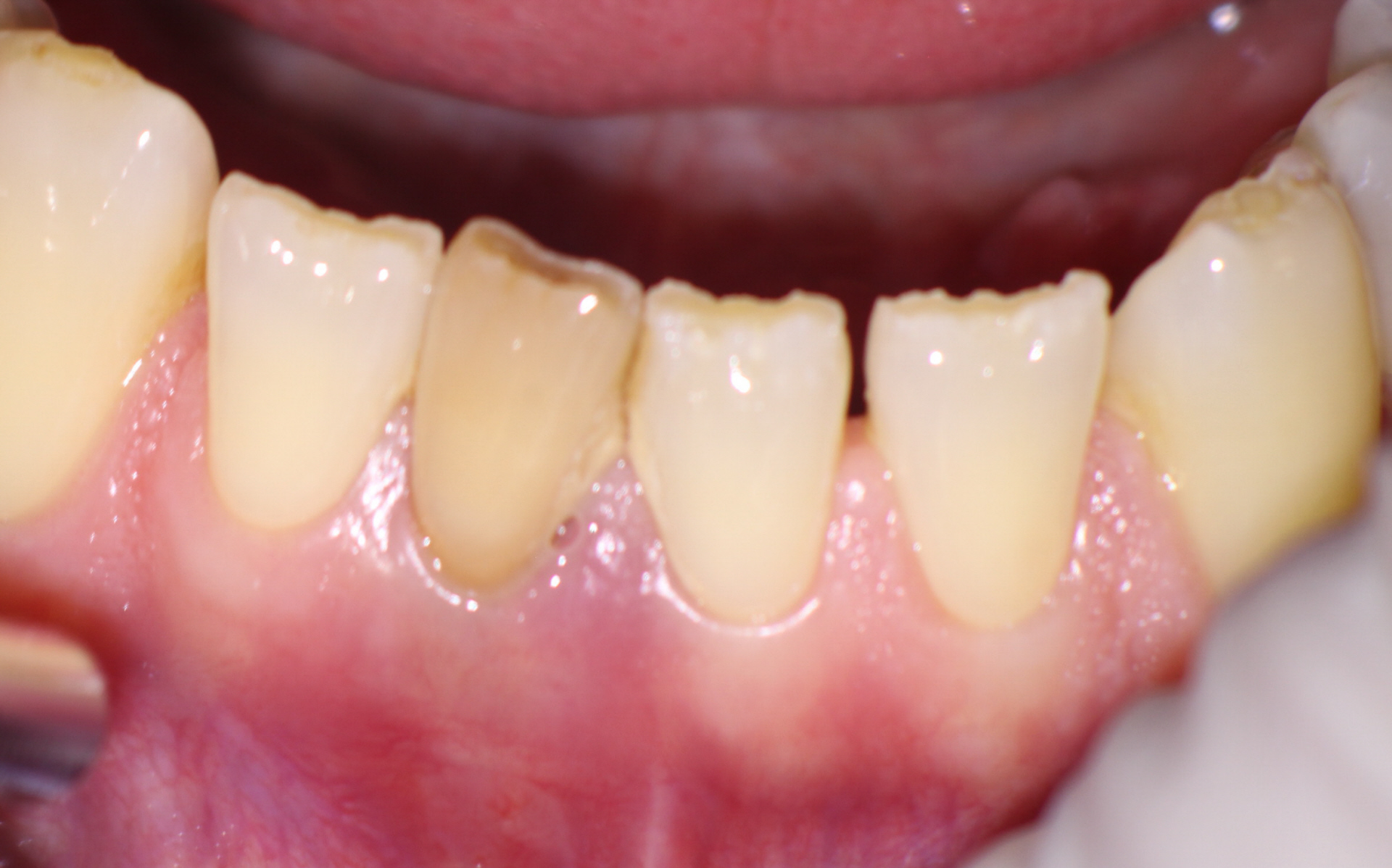
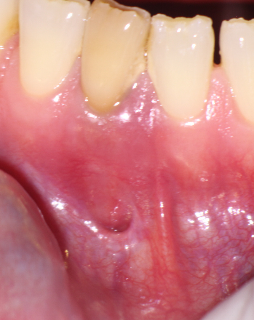
Conventional and cbCT imaging showed complete bone loss around the entire periphery of the root in 360 degrees and hopeless prognosis. PA imaging showed what appeared to be a sliver of radio-opaque material on the mesial side of the root.
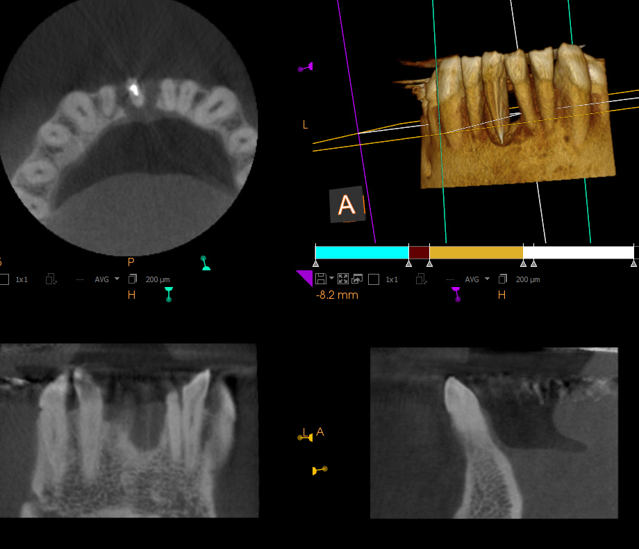
I informed the Referring Dentist that in my opinion the problem was not primarily Endodontic in etiology and that the radiolucency suggested a diagnosis of cemental tear. This resulted in breakdown of Periodontal support from a Periodontal rather than Endodontic problem.
The tooth was subsequently extracted by the Referring Dentist. However, a post extraction radiograph revealed persistence of retained cementum, left behind after the extraction.
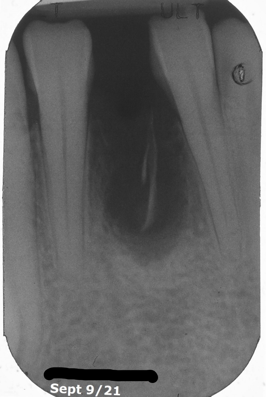
Figures 3 : Referrial’s Post extraction image
Cementum remains. Currette now or remove cementum as part of a regenerative procedure to prepare the site for bridge or implant?
At this point we consulted a Periodontist as to whether the site could be used for placement of an implant and how to best manage any subsequent surgical procedures necessary. Would it be feasible to graft the area sufficiently to allow for placement of an implant? And should we remove the retained cementum now or at the time of possible ridge augmentation?
We agreed that the best way to manage the case was to schedule exploratory surgery of the area by the Periodontist in order to:
(1) Remove the retained cementum and
(2) assess whether ridge augmentation procedures could be performed that would allow implant placement. Should the site (a) not be suitable for an implant or (b) if the patient preferred a different restorative solution (3 unit bridge), there still would be need for augmentation of the socket for proper aesthetics, but perhaps not to the point where it would need to support an implant.
The patient was referred back to the referring Dentist with a copy of the referral letter sent to the Periodontist.
For more information about Cemental Tears – look it up in the EndoFiles Cabinet and search for Author “Lin”, who seems to have done a lot of work in this area.
