Furcal Lesion Heals Nicely
Patients are sometimes referred with furcal radiolucencies or furcal involvement that may or may not be of Endodontic etiology. The question always is: How much of the furcal problem is Endo related and how much is Perio? We know that furcal bone CAN be completely grown back IF the problem is Endodontically related. (Something our Perio colleagues can only dream about!). The decision to commence Endodontic treatment on a furcally involved tooth is complex.
The first thing we need to establish is:
(1) Pulpal necrosis
The pulp MUST be necrotic, at least in the area that communicates with the periodontal breakdown. A finding of normal pulp responses suggests that any furcal or bony breakdown is unrelated to Endo and that Endodontic treatment would be an expensive waste of time and money. Endo won’t help.
(2) The prevalence of Periodontal disease in the rest of the mouth.
Is this an isolated problem, not consistent with excellent periodontal status in the rest of the mouth? Or does the patient have generalized horizontal bone loss and/or chronic Perio disease? If no bone loss is occurring in other areas of the mouth, then it is POSSIBLE that lateral or furcal anatomy could be leaking necrotic pulp products into the involved area, causing breakdown of the supporting attachment apparatus. In other words, excellent Perio in other areas of the mouth, combined with a pulpal finding of necrosis suggests that Endodontic treatment MIGHT improve the situation. There are no guarantees.
(3) Successful fill of lateral anatomy demands that irrigants and medicaments have adequate contact time with the chamber/canal space to work on any tissue that may be in this lateral anatomy. Single appointment, fast Endo treatment will be unlikely to give these solutions and medicaments the time necessary to digest the tissues enough to allow patency and to accept any filling materials, whether it be gutta percha, sealer or a combination of both.
When presenting treatment plan options to patients in such cases, they must understand that the process may be slow and protracted. Treatment will involve multiple appointments, application of irrigants and medicaments and monitoring of the progress of the case until positive results become visible. In some cases this may be rapid resolution of draining furcally related sinuses or closure of deep pockets. In other cases, teeth are temporized until there is evidence of bone fill, which usually takes between 3-6 months to be visible on conventional radiographs. Clinicians involved with treatment of the case MUST provide proper, secure, durable, sealed temporization to ensure asepsis and prevent contamination.
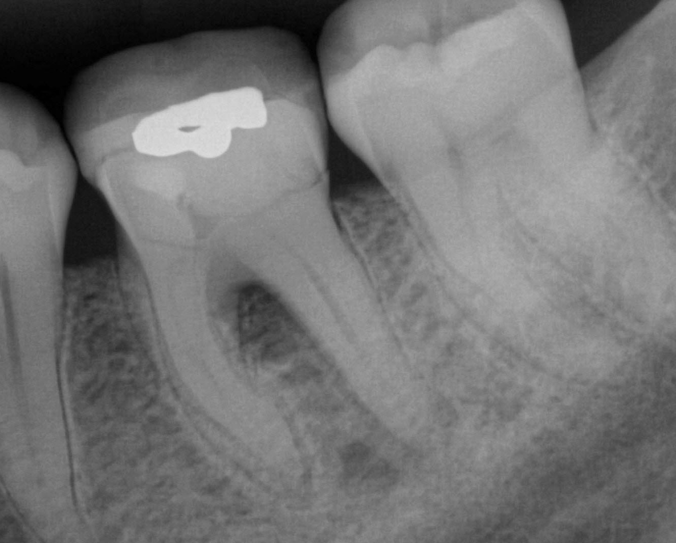
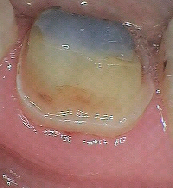
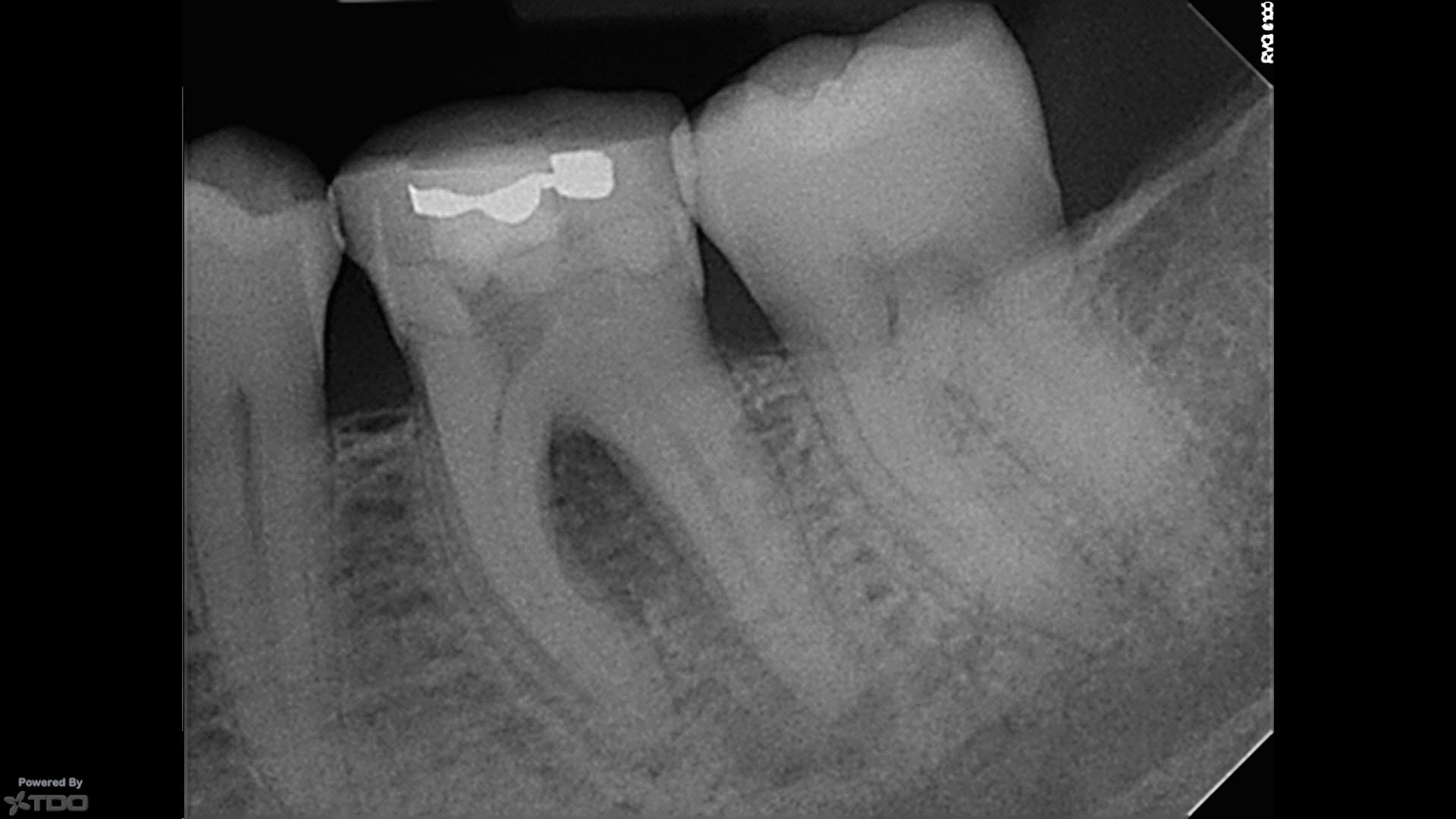
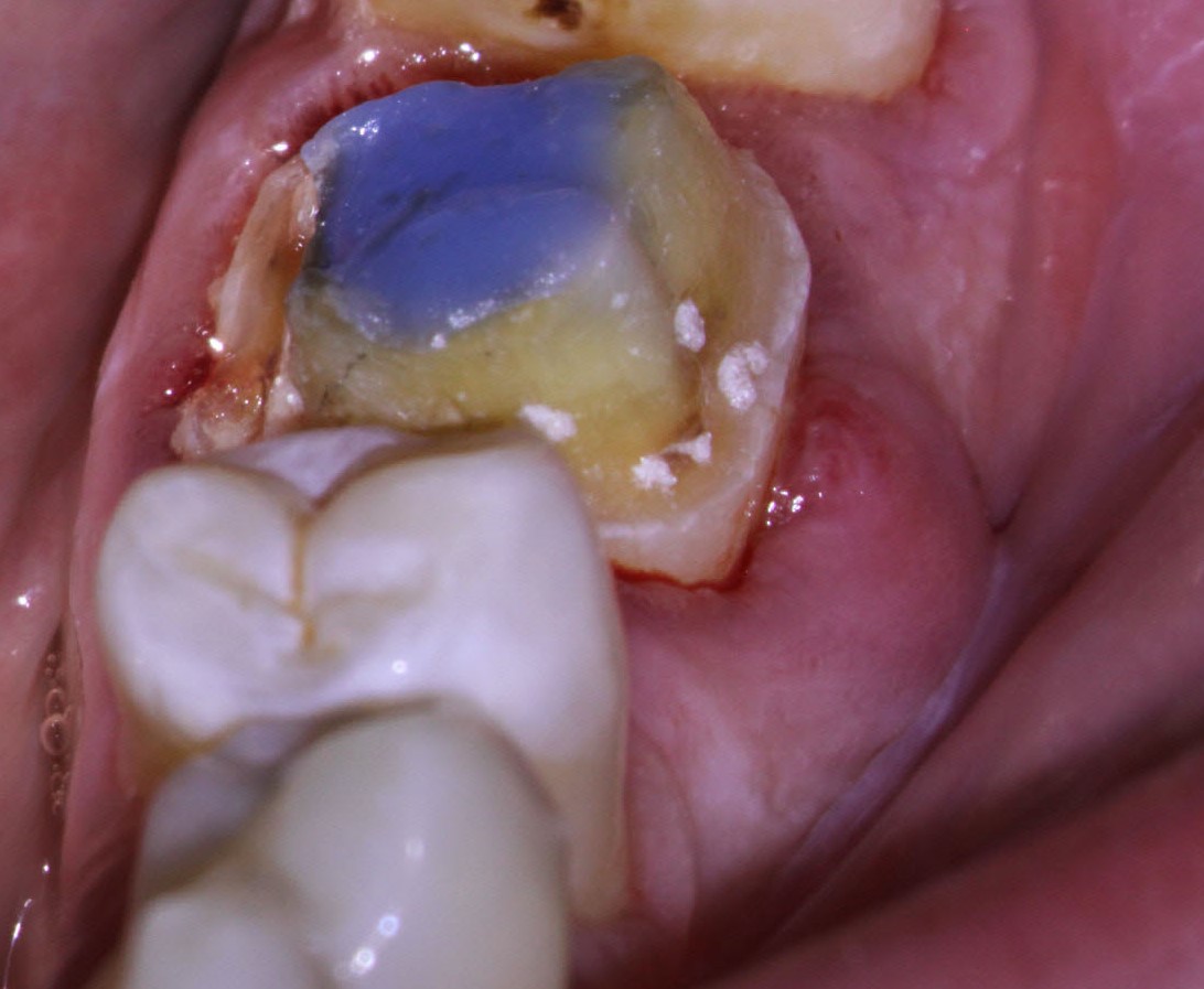
Tooth on presentation in my office. Access had been attempted by the referring Dentist and sealed with colored composite.
In this case, the referring Dentist (an excellent GP clinician) started the endo on tooth #36. His referral note was excellent and comprehensive.
“The patient had caries under the crown. The crown was removed, and the caries excavated . I determined that the tooth was salvageable. A temp crown was made and the patient was appointed to do the Endo. Upon access, I had trouble finding all 3 (or 4) canals and was only able to find one. I am referring the case to you. I noted the furcation radiolucency with subsequent Perio probing that was not present in the radiograph in the patient’s previous radiographs. (Important history!) He suspected that the furcal involvement was of endodontic origin creating an endo-perio lesion.” Great note and good call!!
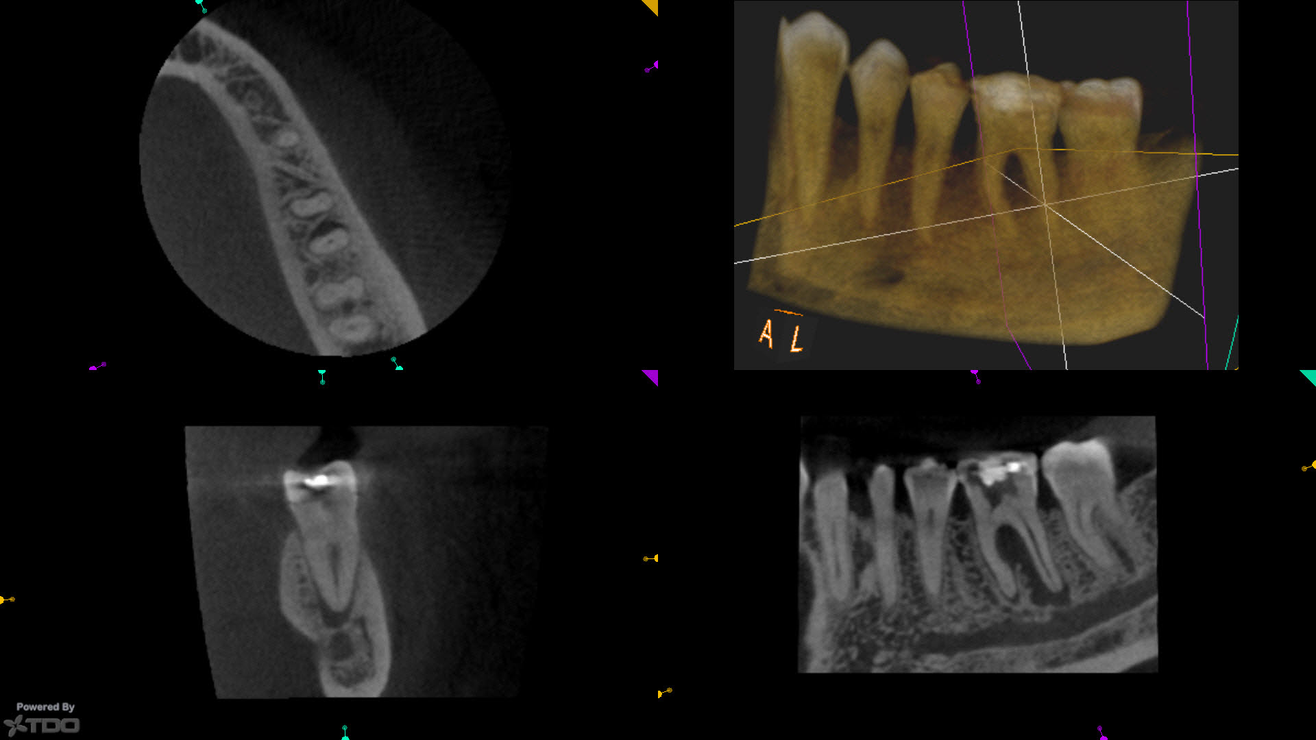
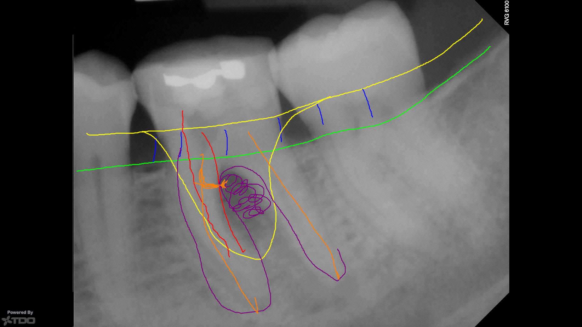
Diagrams and annotated illustrations help explain the nature of the problem and treatment goals to patients.
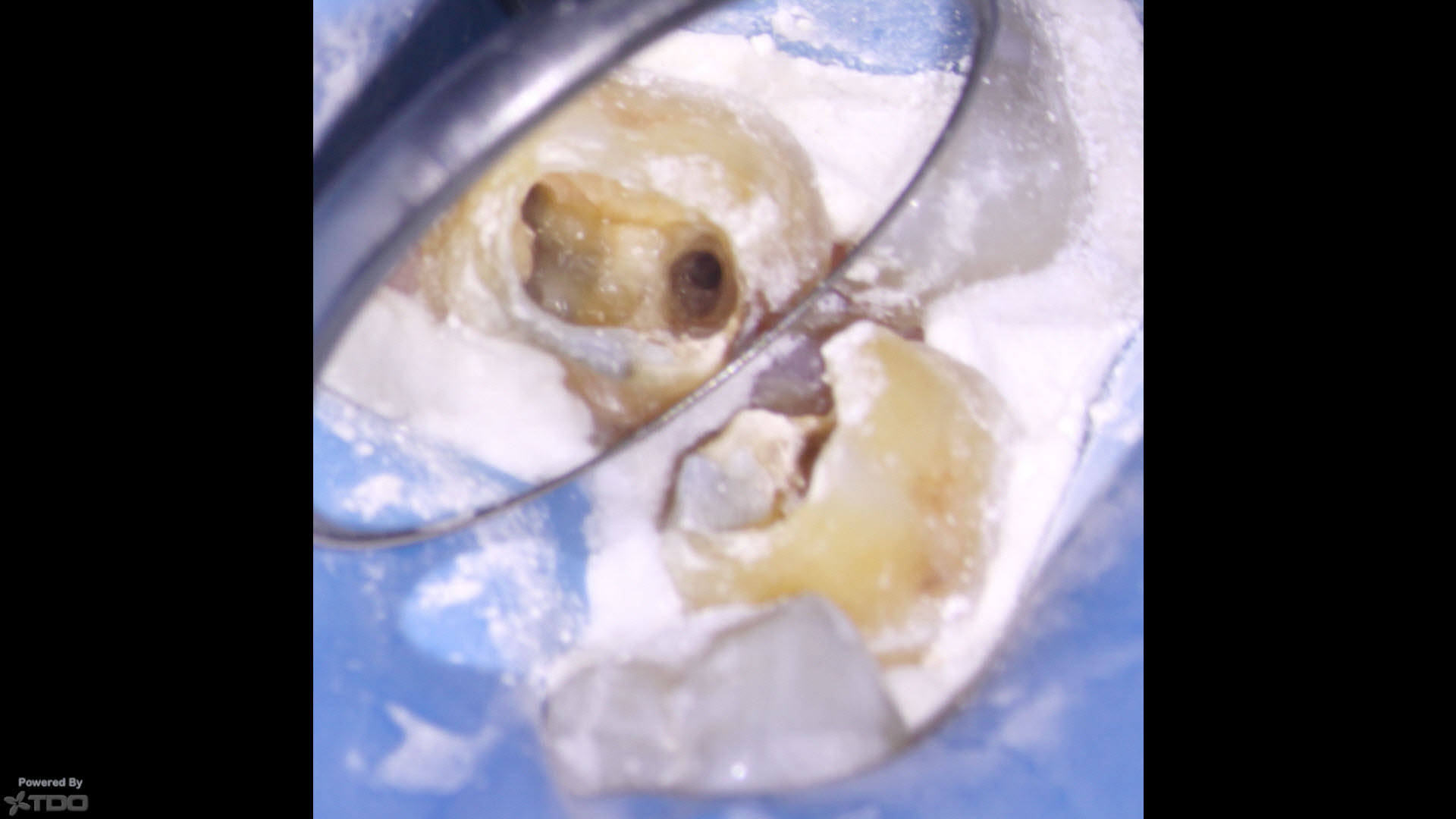
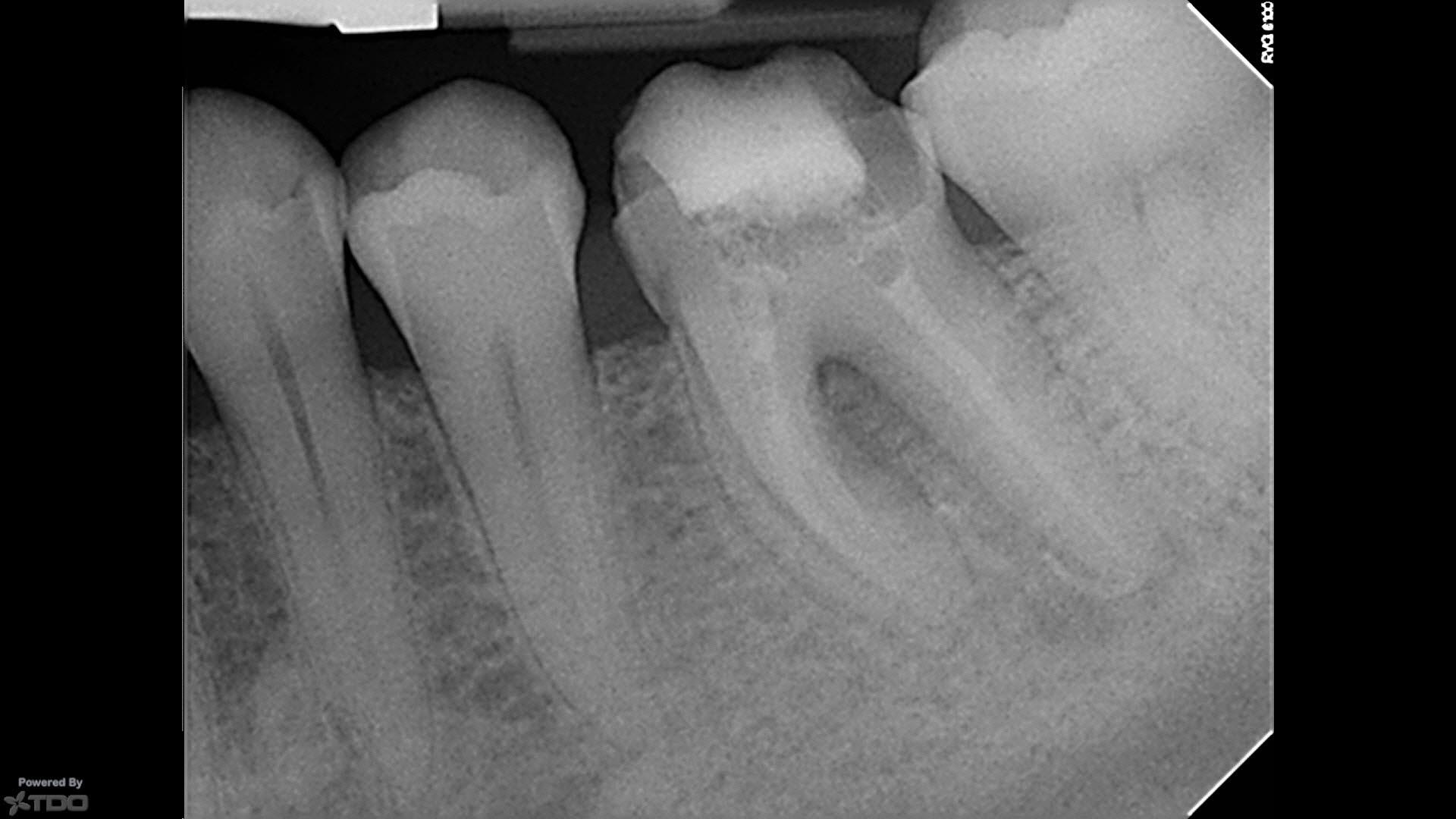
Clean and shape of the canals and Ca(OH)2 medication.
At 4 months and we began to see signs of furcal regeneration.
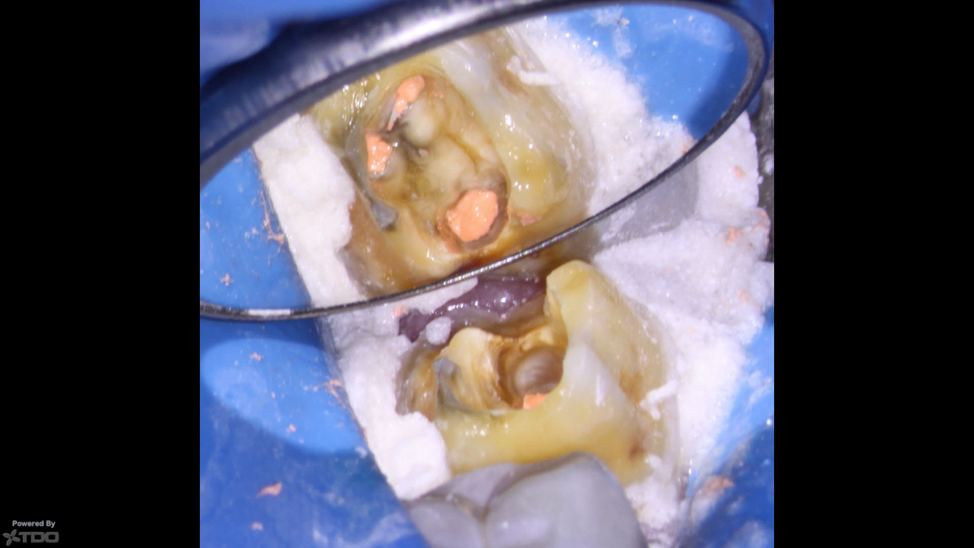
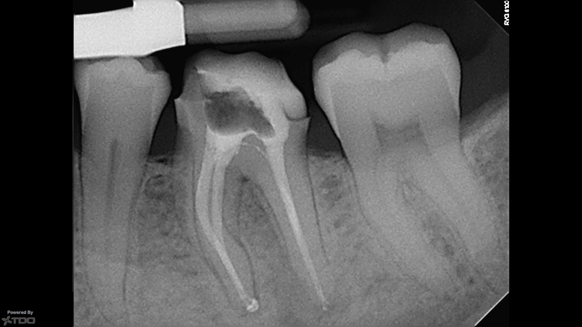
Canal system obturation was performed with instructions to immediately return to the dentist for final restoration.
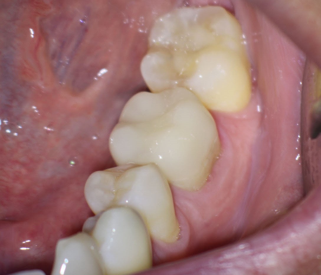
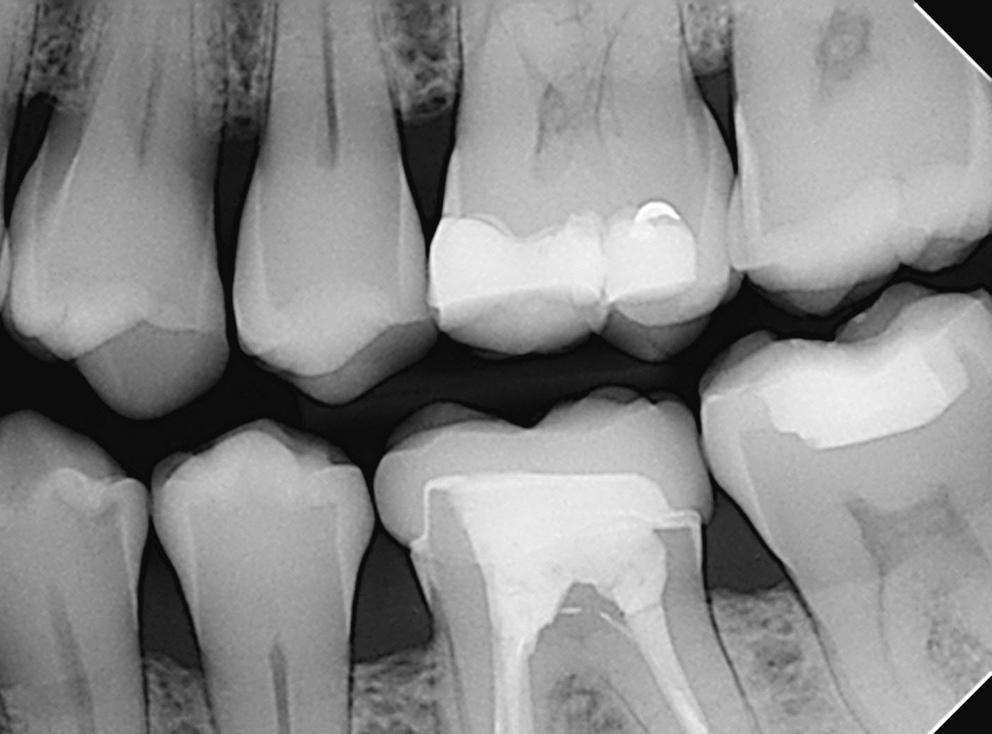
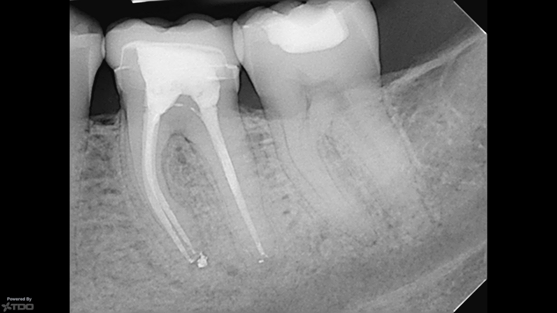
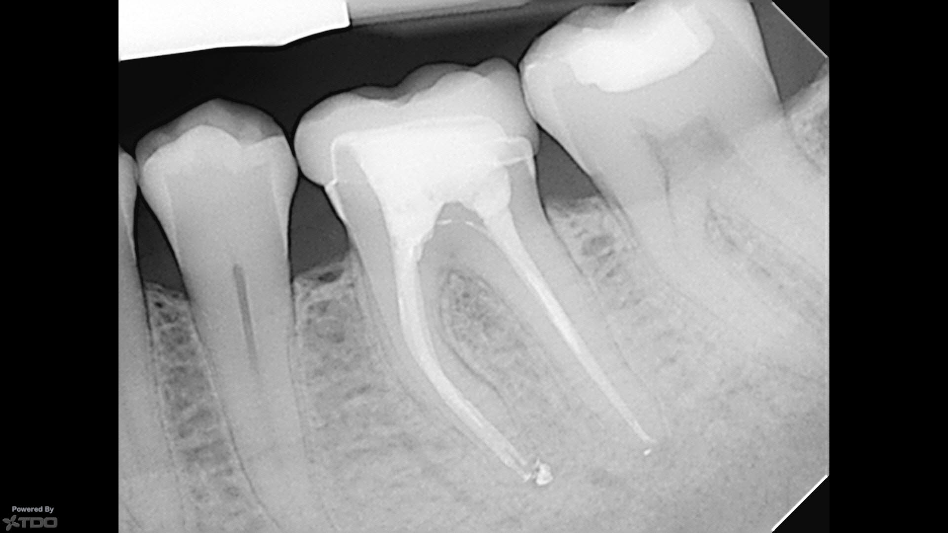
The tooth was then beautifully restored. The BW and last 2 periapicals show full furcal regeneration and a nicely restored tooth.
