Multiple Foramina
Although cone beam tomography has wonderful imaging capabilities, treatment of what appears to be relatively normal canal anatomy can result in some interesting final results.
In our move to mechanization of canal instrumentation through the use of Rotary nickel titanium engine driven instruments, we sometimes forget the need to explore the apical extents of canals with hand files or become frustrated when the anatomy is challenging.
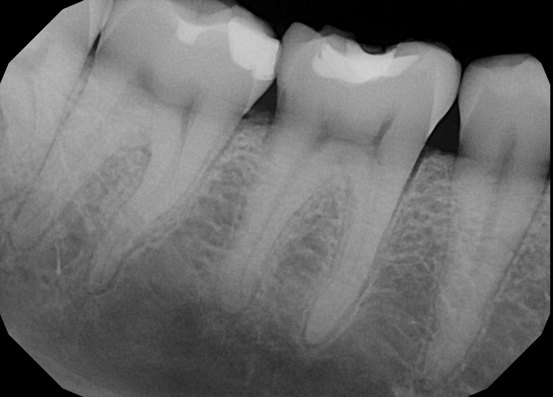
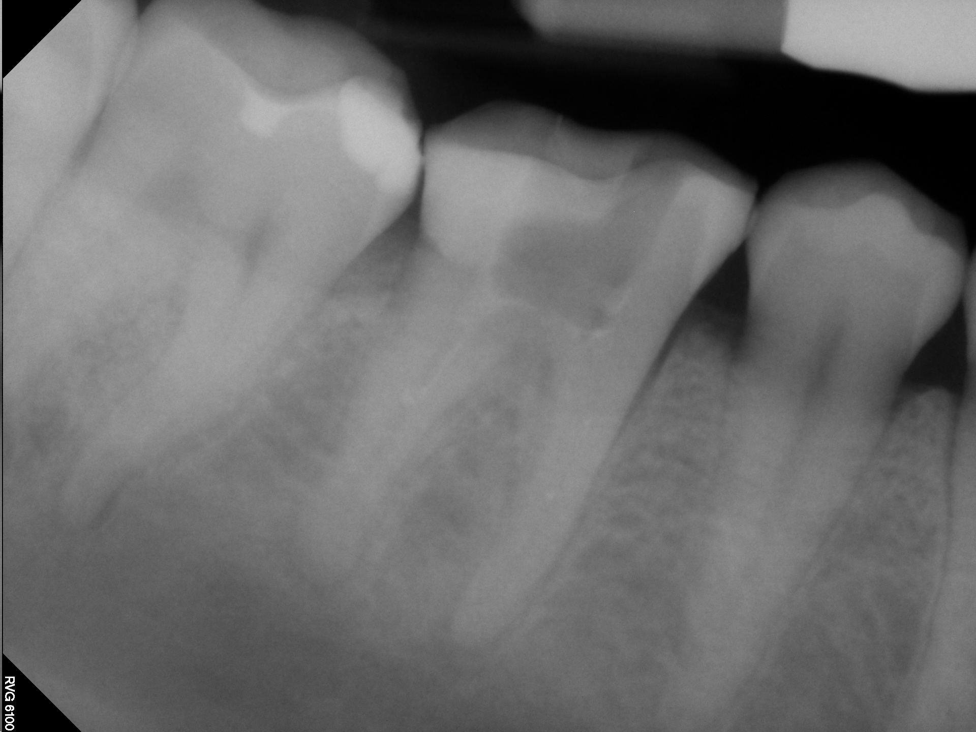
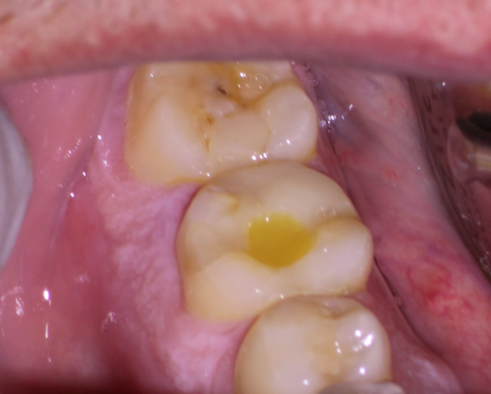
This case was referred to me after access and Ca(OH)2 placement because the referring Dentist thought the “apex was calcified”. This is a common theme among referrals who simply do not understand that it is impossible for the “apex to be calcified” because:
(1) The coronal aspect (the canals and pulp chamber CANNOT be vital without communication of the pulp with the periapical tissue.) These tissues cannot be “calcified”, otherwise the pulp chamber tissue could not survive in a vital state.
(2) Endodontists know that with only a few exceptions (severe abfractions, and buccal and palatally restoratively induced calcifications) pulp tissue generally calcifies in a coronal to apical direction. The LAST place to calcify in almost all cases is the apex. This is why we often see completely obliterated chambers/canals in anterior tooth trauma cases but with apical radiolucent findings caused by a small bit of remaining non-vital apical pulp.
We are frequently in a rush to complete the case and because of this sometimes the anatomy is not filled as well as we would like. In this particular case I detected multiple foramina at the apices with the use of very small hand files and tried my best to maintain patency in more than one direction. This is extremely difficult because this is so far down the canal and failure to place the instrument into the access at the proper angle can result in either instrument fracture or ledging of the anatomy that was previously found.
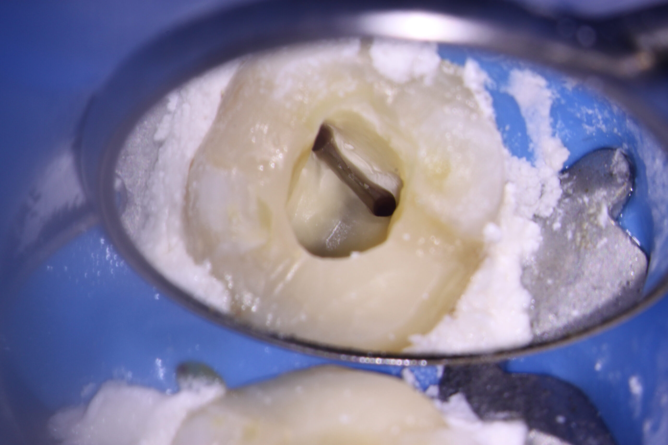
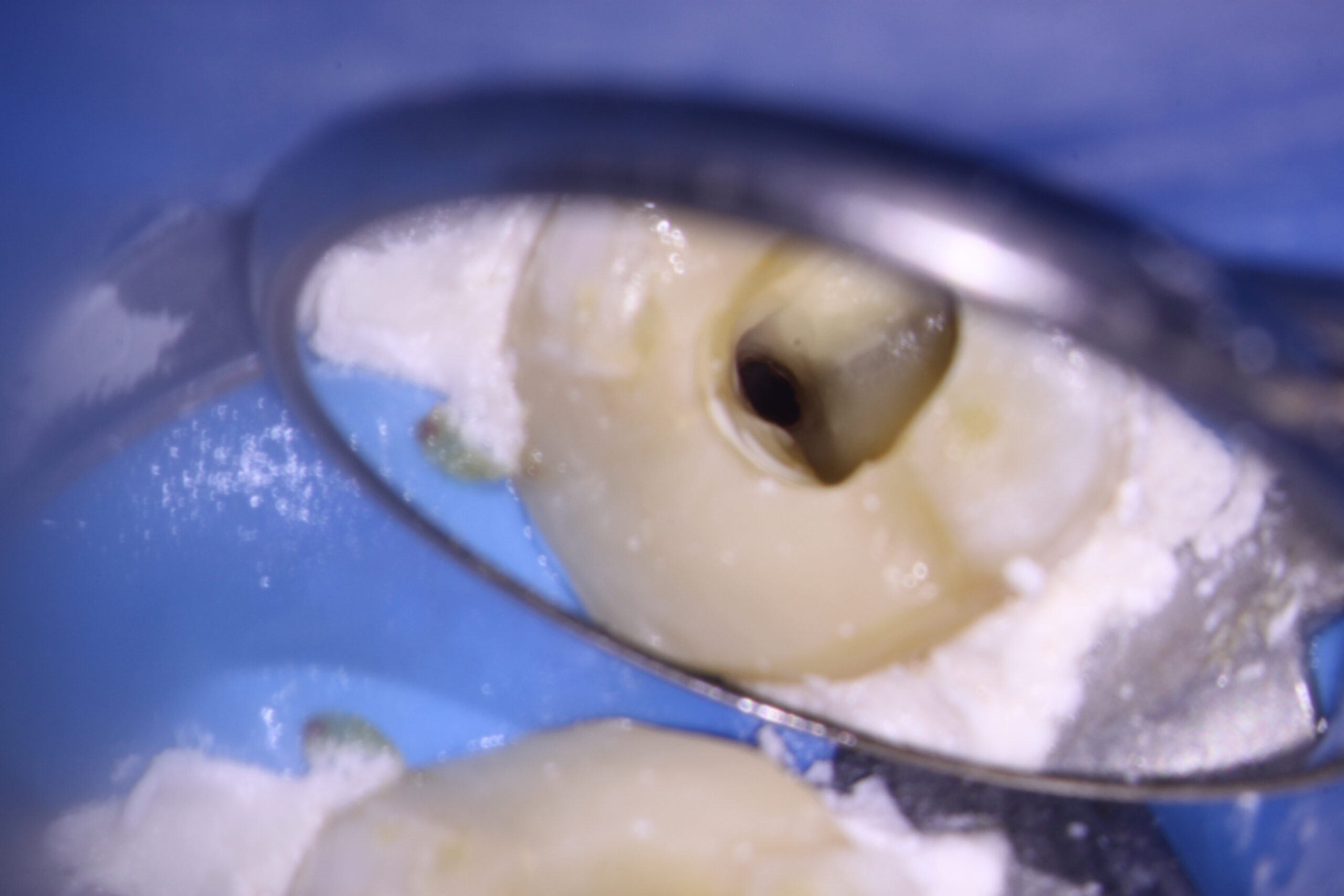
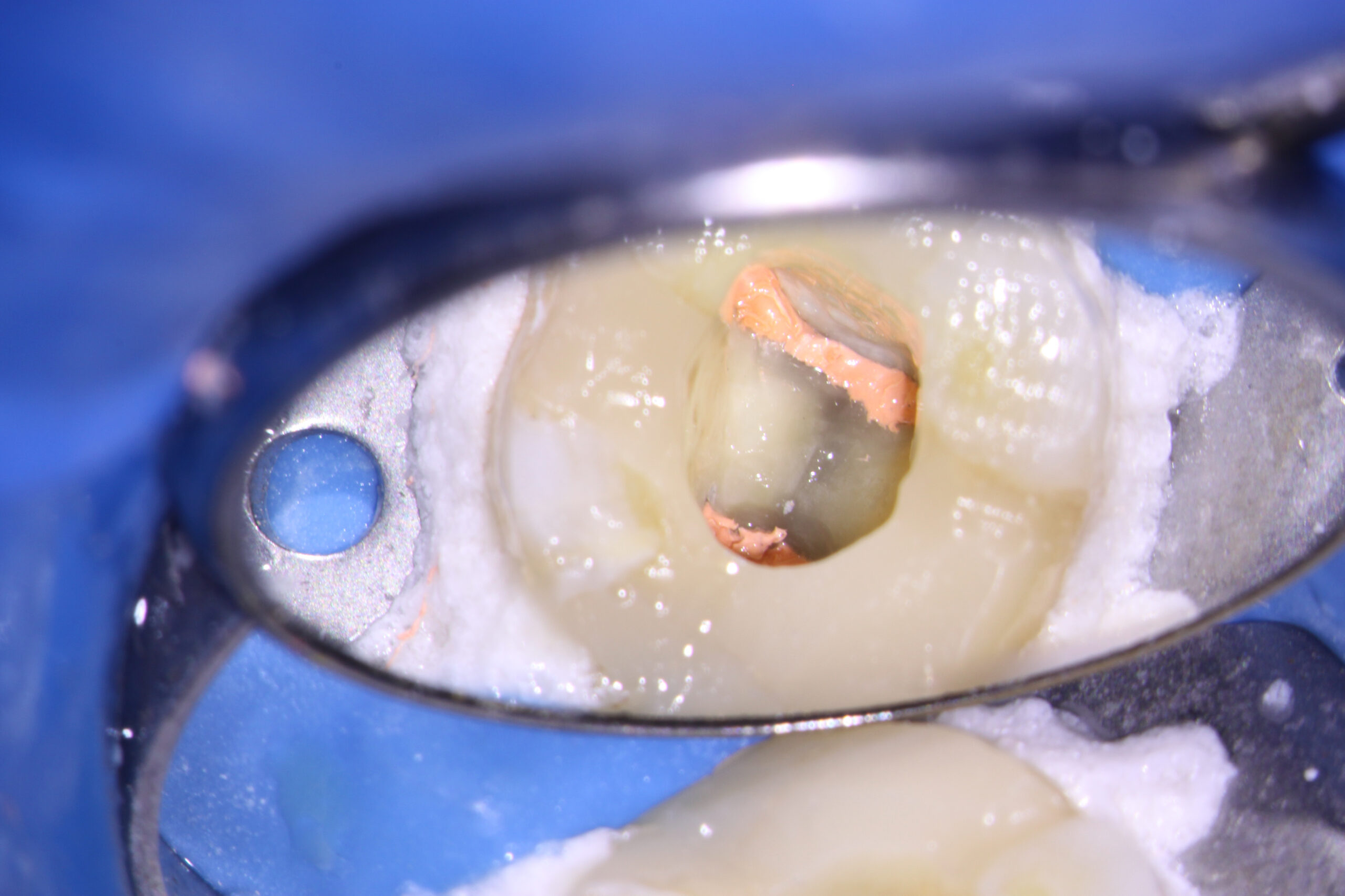
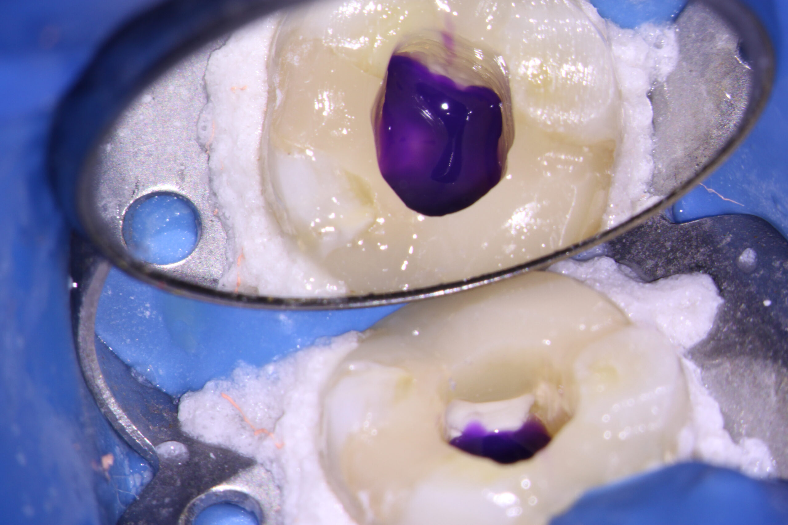
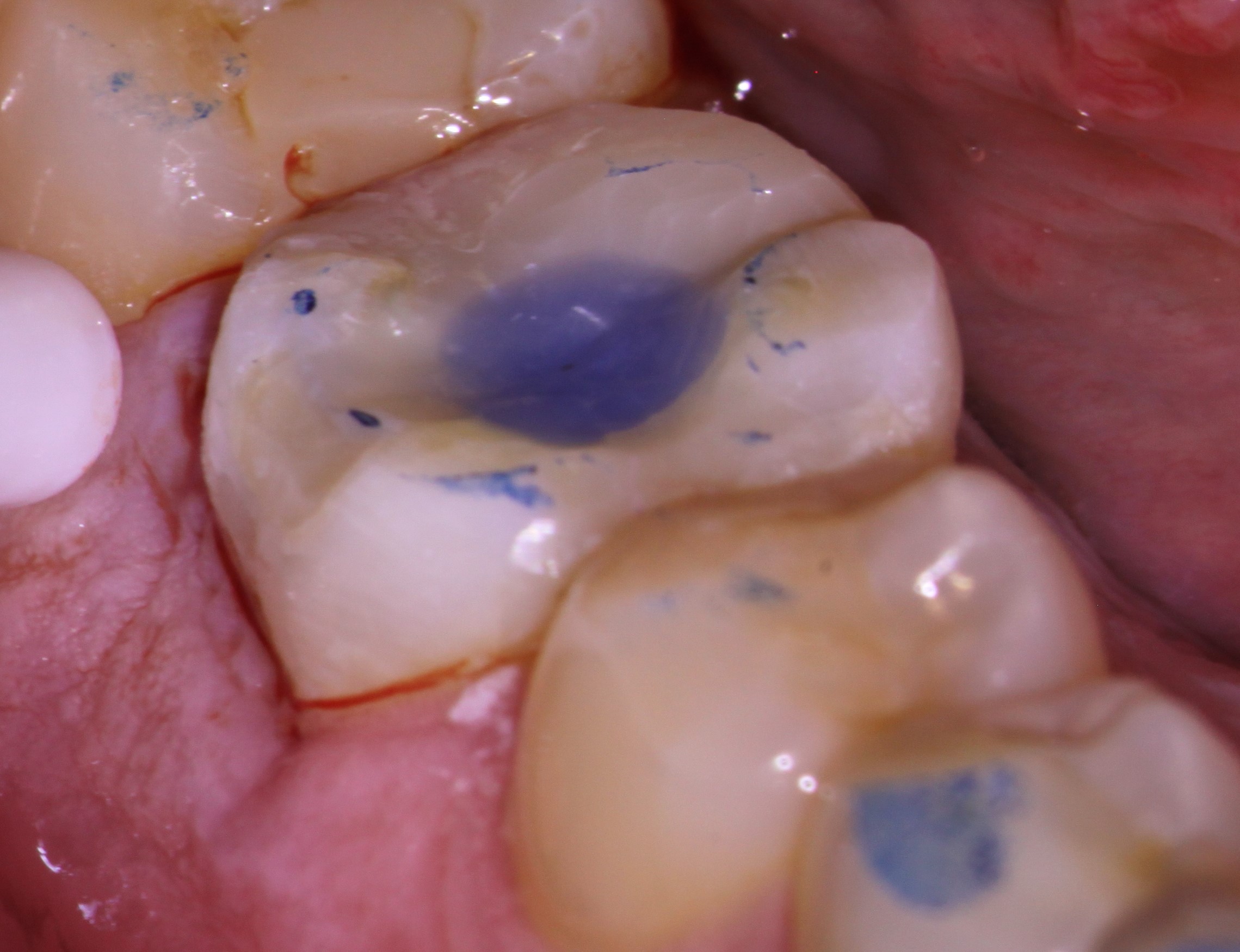
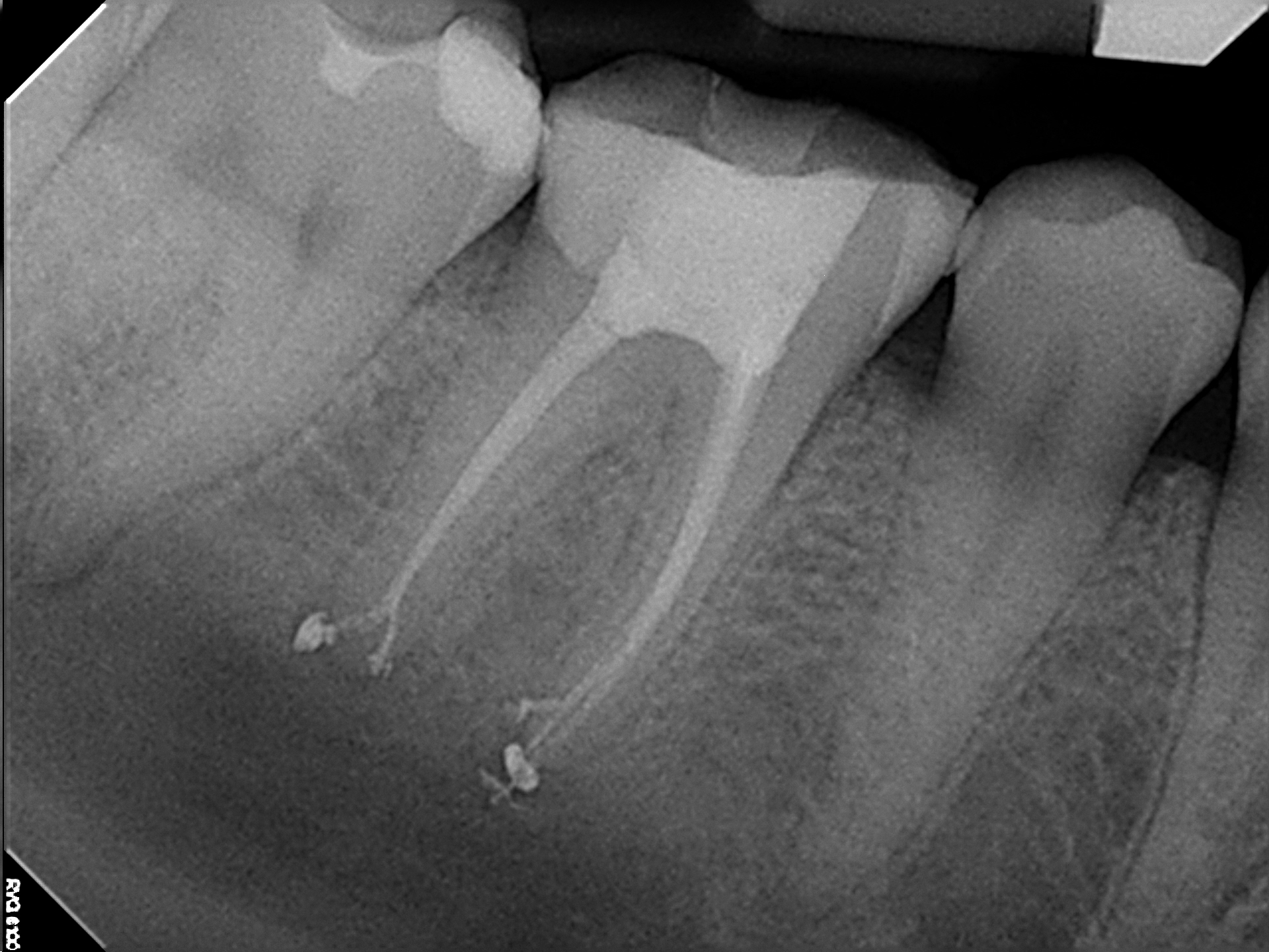
I was extremely happy with the final result and the fact that the referring dentist allowed me to place the orifice bond as well as the core filling . The patient was referred back to the referring dentist knowing but the prognosis for this tooth was excellent.
