Lateral resorption Affects Preparation Technique
This 45 year old patient was sent to me for examination regarding diffuse radiating symptoms in the anterior mandible. The referral’s note said:
Please examine patient to check lower left. We think #32 is the problem. Patient feels pain radiating whole lower left side. Pain is on/off, comes in waves. When it hurts it really hurts. We referred patient to to an Oral Surgeon in 2005 to extract #31. The patient remembers it as being a tough extraction.”
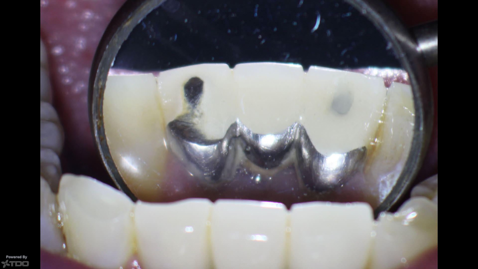
The patient was relatively asymptomatic upon presentation. Clinical tests showed positive response to percussion and biting pressure on the #32 abutment of the bridge but this was difficult to interpret because of the connection to the other healthy abutment. Palpation of the buccal gingival area of the retained root was normal. Chewing was positive as well, but again, this was somewhat ambiguous due to the attachment to the other bridge abutment. Pulp test showed no responses to cold or hot in #32 pulp tests, but again, these results could be false because they may not yield reliable results when they were performed through a casting. Gingival probings were normal.
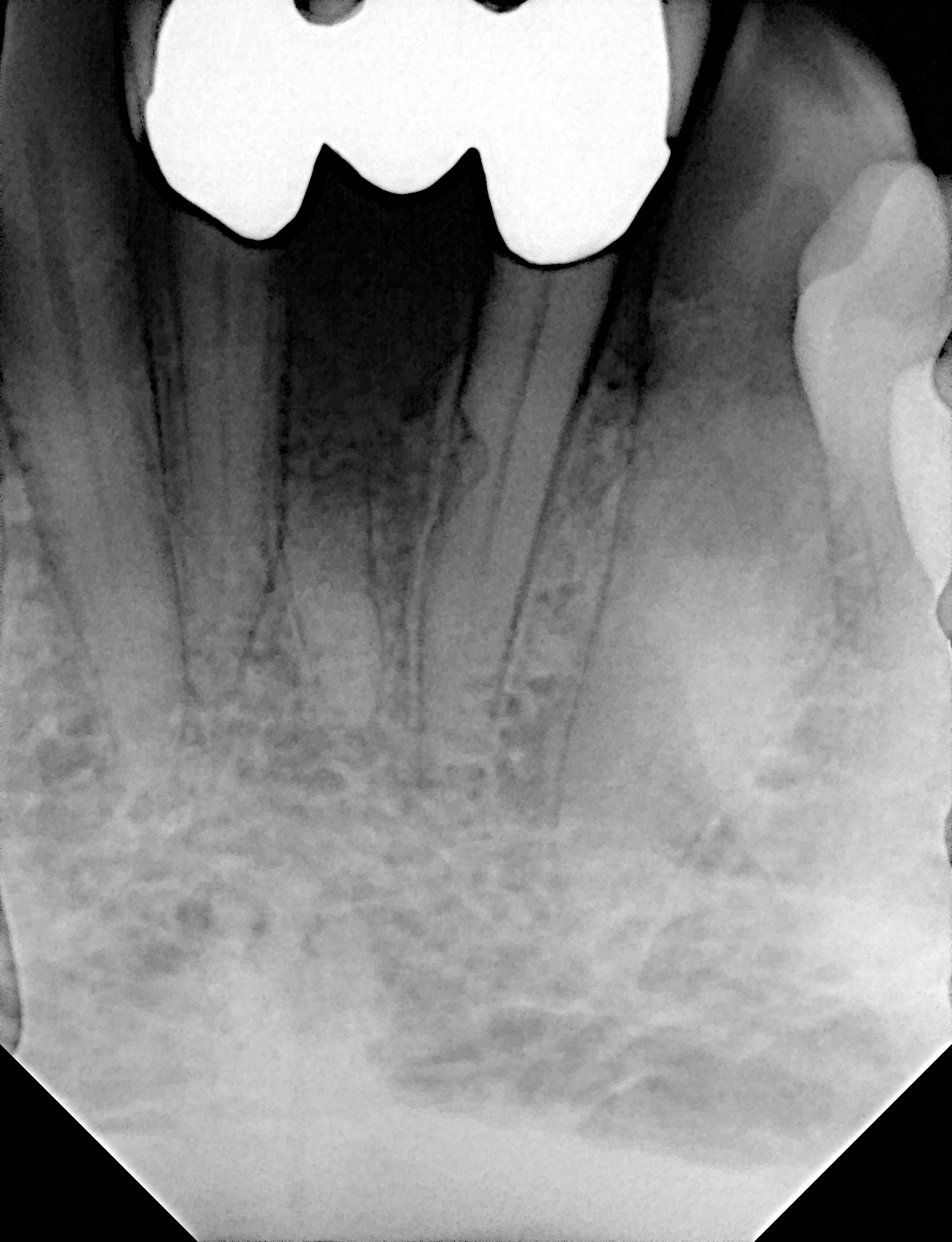
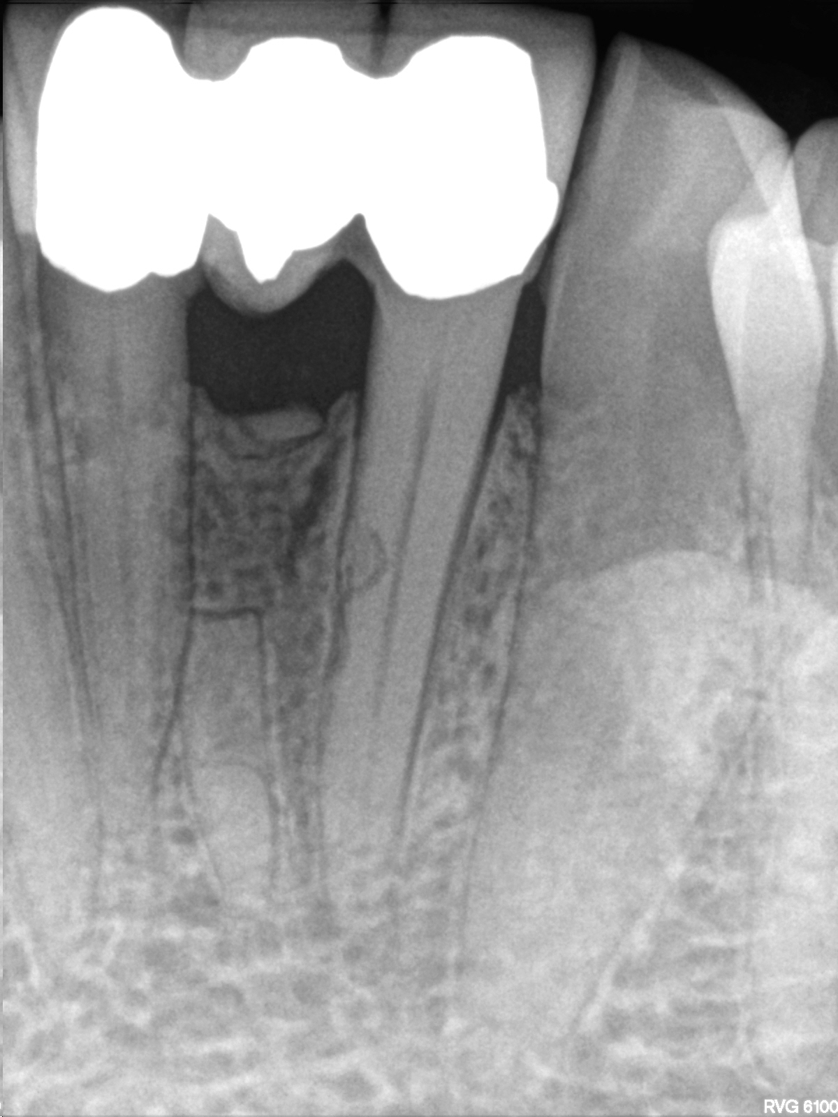
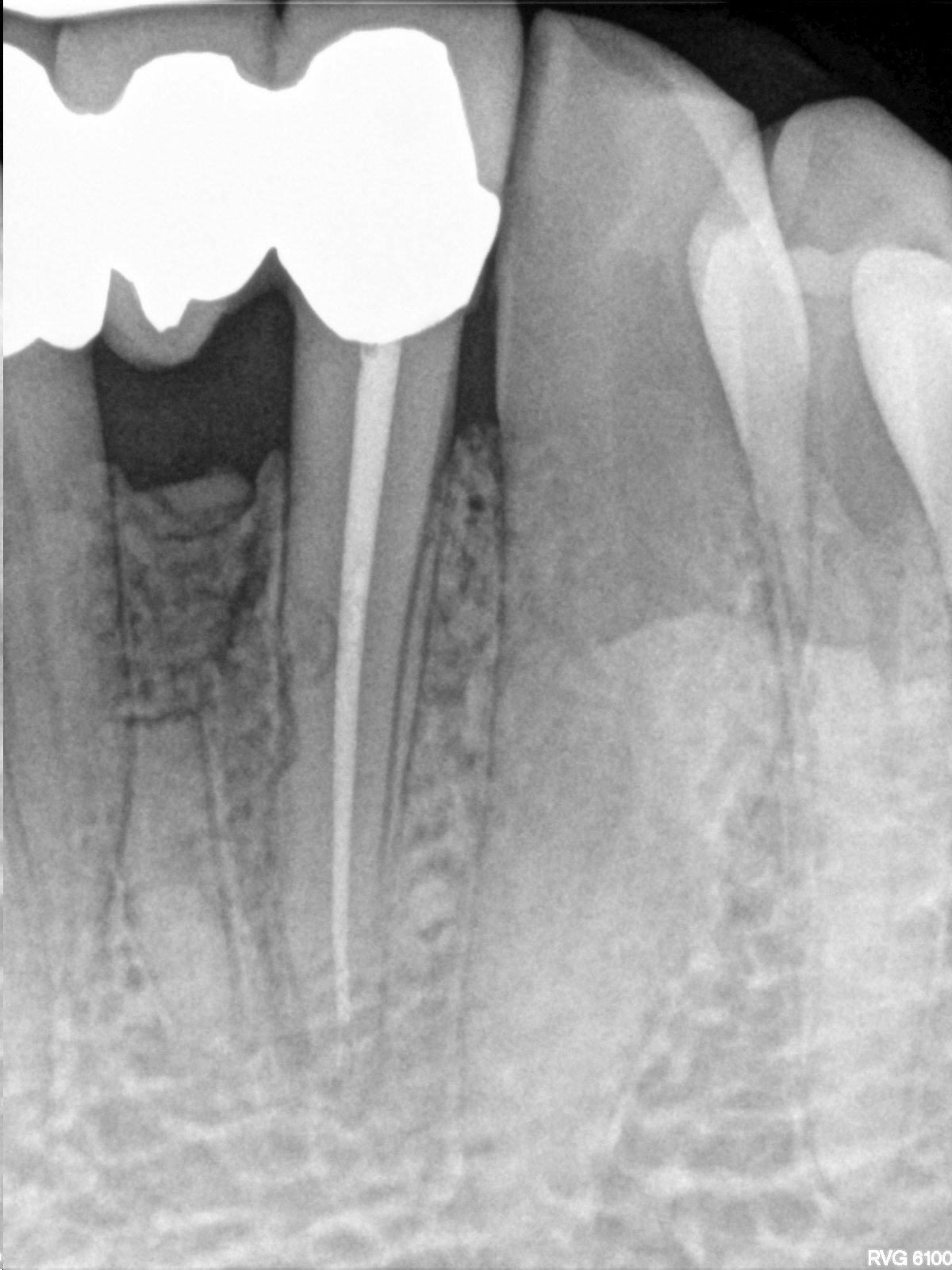
Periapical radiography showed the presence of a 3 unit bridge #41 -32 with good margins with evidence of a retained root tip in the position of tooth #31. I thought this unusual considering that the original extraction was performed by an Oral Surgeon, who usually don’t leave root tips in place. There were no unusual radiolucent findings associated with the root tip. of #31. I was not sure of the reason for the original extraction. (Trauma?)
Cone beam tomography revealed a small radiolucent area at the apex of tooth #32. In addition to the retained root tip, I noted that there was an area of root resorption on the mesial midroot aspect of #32. I was not sure whether this may have caused by the effects of the original trauma, attempts to remove the central incisor during extraction or whether it was simply idiopathic. In any case, any Endodontic treatment that was being considered had to take this area of minimal dentin into consideration during canal preparation.
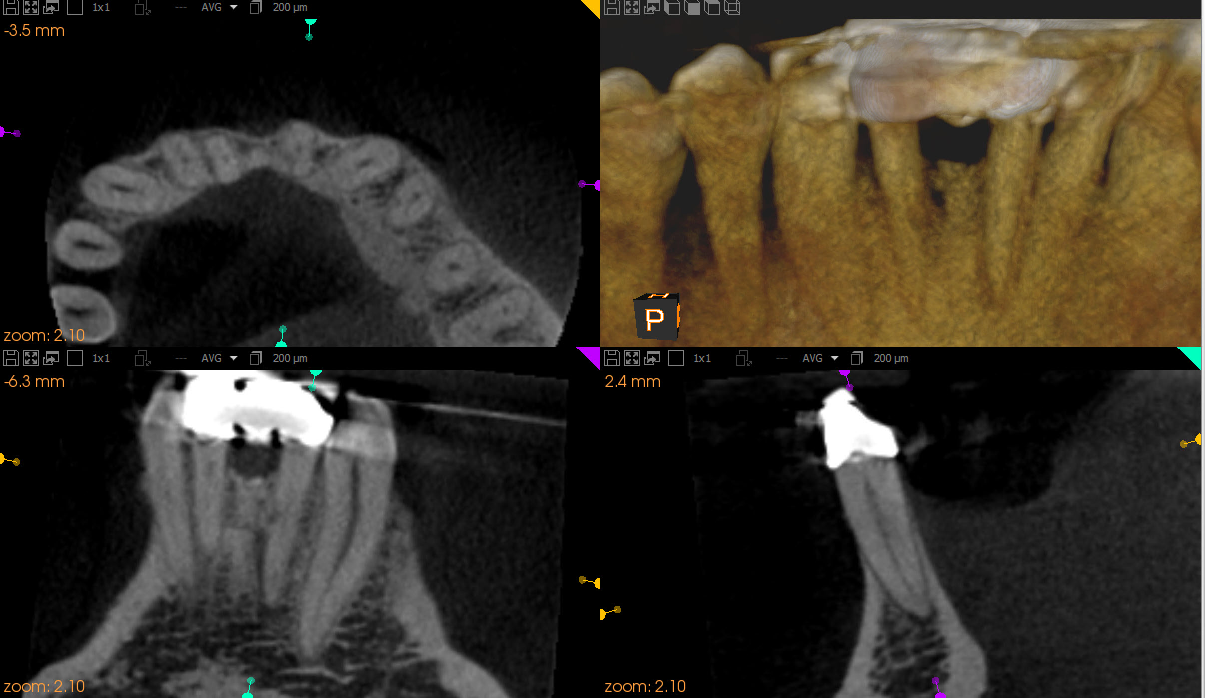
I explained to the patient that there was radiographic evidence that the pulp of this tooth may have undergone necrosis and that in order for us to rule out the tooth as a source of his discomfort we would need to first (a) do a cavity test and if so (b) endodontically treat # 32 through the Crown. I also mentioned that I noted an unusual area of mesial resorption and there was a small chance that preparation of the canal could lead to exposure of this lateral area but that every effort would be made to keep the access and canal preparation conservative and avoid communication with this reserved area.
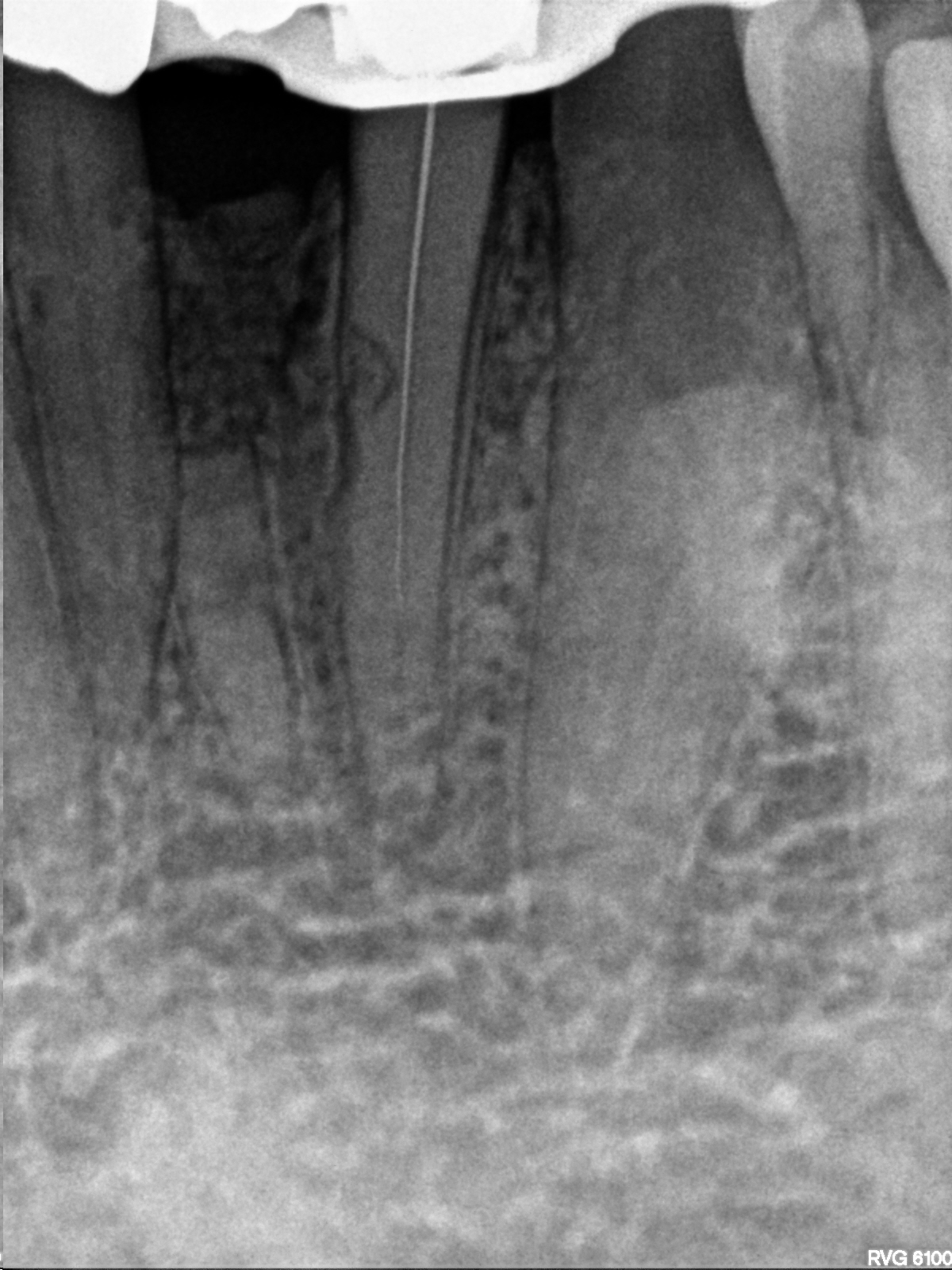
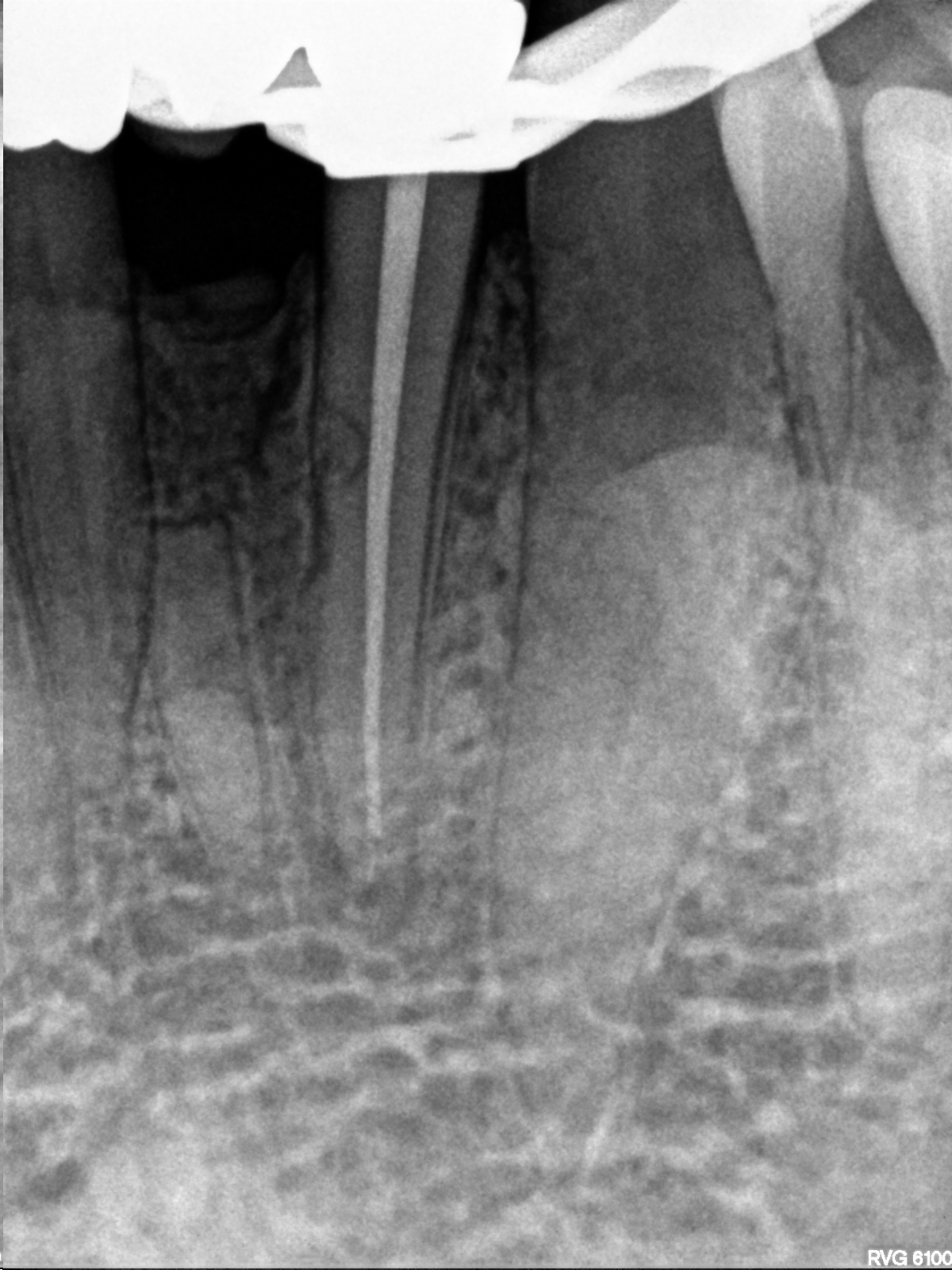
A rubber dam was placed and access was made through the Crown without anesthesia. The pulp was found to be non responsive and when we accessed the chamber we confirmed pulpal necrosis. The canal was cleaned and shaped using minimal preparation techniques (Edge rotary Ni-Ti files) and obturated with F-M gutta percha cones and vertical compaction of warm gutta percha. The access was closed with composite. It was especially important for us to be conservative with canal shaping to prevent the possible communication with this resort area and possible extrusion of filling materials into the lateral side of the root. Fortunately, the canal was straight and access was quite easily performed. The patient’s symptoms resolved with Endodontic treatment.
