Cavity Test Aids in Proper Diagnosis
A 26 year old female patient was referred to me for consideration of the mandibular Central Incisors The referring dentist had seen a periapical radiolucent area associated with the apex of these two teeth (mostly with tooth #31) and was unsure of the diagnosis. The patient was asymptomatic and had a history of orthodontic treatment . There was no history of trauma to the area. She came in with lingual arch wire, placed to help maintain position of these orthodontically treated teeth. The crown of tooth #41 appeared to be slightly discolored, though the difference in color was small. There also appeared to be some apical resorption at the the root tip of #31 that was typical of pathology associated with teeth that had chronic periapical periodontits caused by a necrotic pulp. Percussion palpation and periodontal tests all revealed normal findings
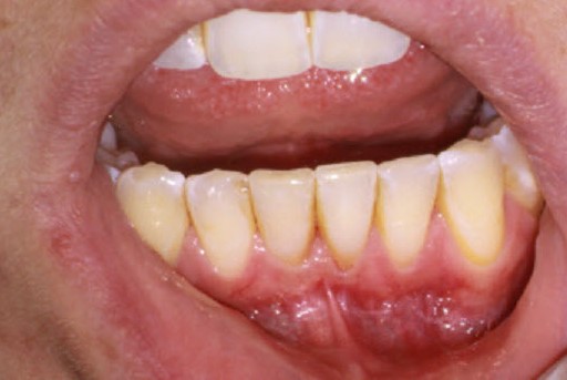
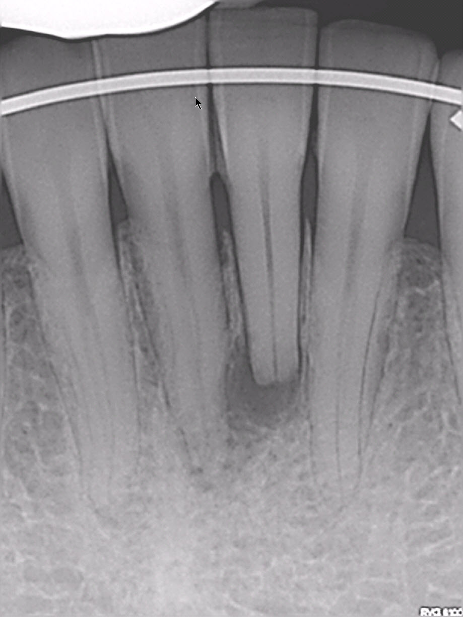
Pulp tests performed on all the anterior mandibular teeth showed normal responses with the exception of #41, which did not respond. This was unusual because the radiolucent finding appeared to be centered at the apex of tooth #31.
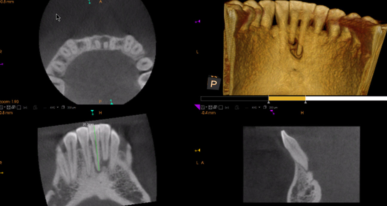
CBCT imaging showed a diffuse radiolucent apical area and bone loss encompassing apices of both #41 and #31 on the lingual side rather than the buccal cortical plate. The bone loss seemed to be centered around tooth #31 but this was not consistent with the presence of normal pulp test responses in this tooth.
I explained my findings to the patient and suggested that a cavity test was in order so we could confirm a definitive diagnosis of pulpal necrosis in #41.
A decision was made to remove the archwire and make access through the lingual aspect of #41 to allow us to do a cavity test. Penetration of the bur through the enamel revealed no response from the patient and the chamber was entered. The pulp was found to be necrotic and working length was easily obtained without the use of local anesthetic. Because the patient was traveling 8 hours by car (one way) to see me, we decided to complete the endodontics in one visit. The access cavity was closed with composite and I instructed the referring dentist to replace the archwire if he felt it was necessary.
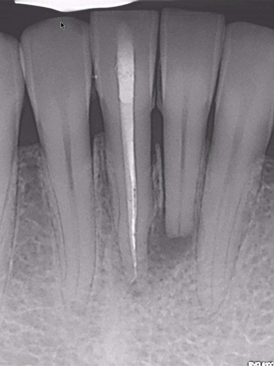
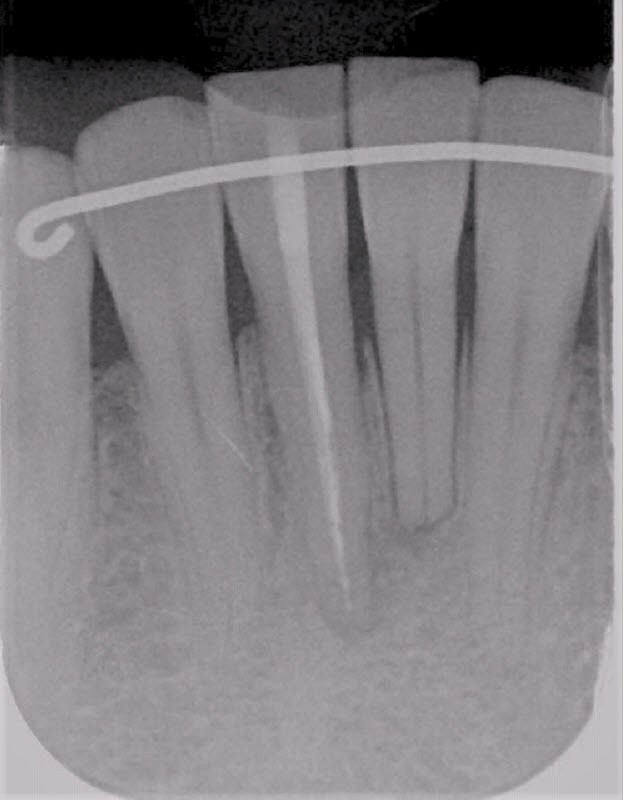
The referring dentist sent a 6 month follow up radiograph (right) which shows good healing around the Endodontically treated tooth and the adjacent #31.
There is a tendency to look at a radiograph, see an associated radiolucent area and assume that the tooth adjacent to the radiolucent finding is the source of the Endodontic problem. This is not always the case and this is a good example of why it is important to perform pulp tests. Where a necrotic tooth is suspected, the cavity test is the final test to ensure the diagnosis is correct. Although this may seem a little unsettling for the patient, ensuring proper diagnosis ensures that the correct tooth is being treated.
