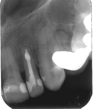
Classic Internal Resorption
In this case of the month we revisit an old case that had been previously accessed and left open. This 45 year old female’s maxillary left lateral incisor had a history of deep composite restorations on both mesial and distal sides. She was Hepatitis B positive and also had a history of orthodontic treatment later in her mid 30s..
The apical third showed a fairly large area of internal canal resorption. The apex shows a classic lesion of endodontic origin associated with a necrotic tooth. The patient was asymptomatic with the exception of a draining sinus on the labial aspect.
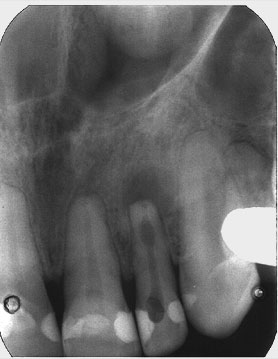
A working length was established used a K file (Size #40). The canal was cleaned and shaped using standard irrigation protocols ( NaOCL 5.25%)) taking care not to enlarge the apical foramen that appeared to have been resorbed as well. ( It is unknown whether the resorption occurred as the result of pathology or Orthododontics) This case was done in a single appointment, without placement of medication such as CaOH. A coarse paper point was used to gauge the foramen size and point of termination.
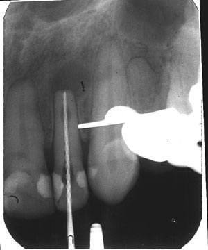
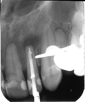
The case was filled with Warm Vertical compaction technique. Post op fill shows good adaptation to the resorbed area and no excess out of the canal space. The case shows good apical control while at the same time filling a large space close to the foramen.
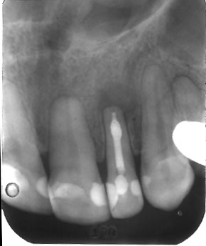
A 4 year recall film shows complete bone fill and healing. Internal resorption cases make up only a small percentage of resorption cases that Endodontists see. Most “internal resorption” cases diagnosed by referring dentists are actually misdiagnosed external resorption.
This is a true internal resorption case with a classic appearance of the resorbed are of the canal. These case respond very well to proper cleaning and shaping although they are not the “Classic canal shape”. Warm vertical obturation with gutta percha is particularly well suited to obturate such cases because warming the material allows it to adapt to these “non-standard” shapes.
