3 Distal Canals Mandibular Molar
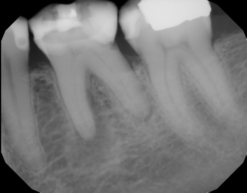
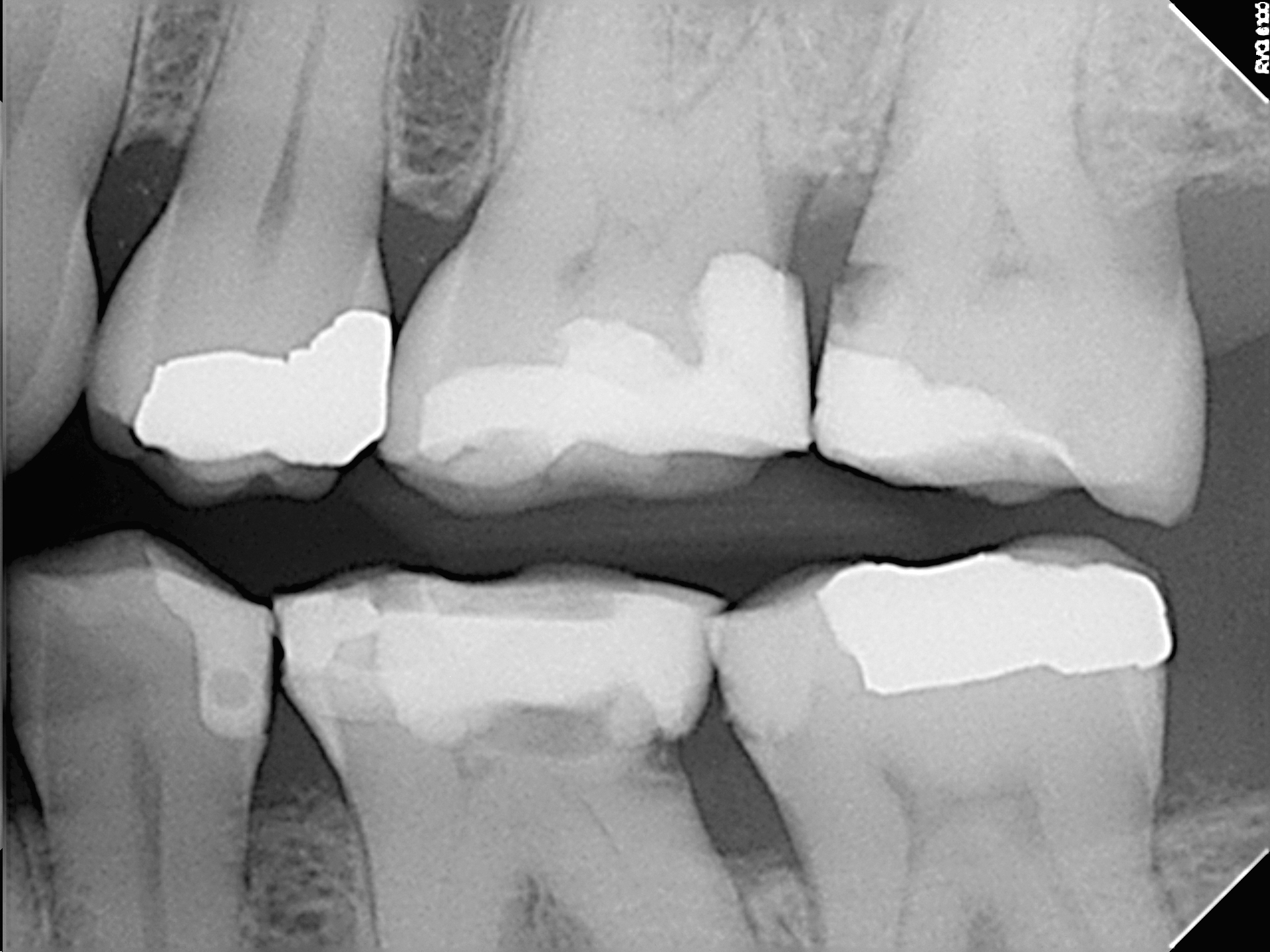
Here is an interesting case that was sent to us . 80% of the cases I see are molars of some type. The distal canals of molars are often extremely variable in their anatomy. While many conditions think of the mesial canals is more challenging, the anatomical variation in the distal canal of these mandibular molars can be extremely variable. In this case the preoperative radiographs didn’t really show very much an we assumed that because the tooth was relatively small , the roots were straight and the anatomy looked fairly typical, that this would be an uncomplicated case.


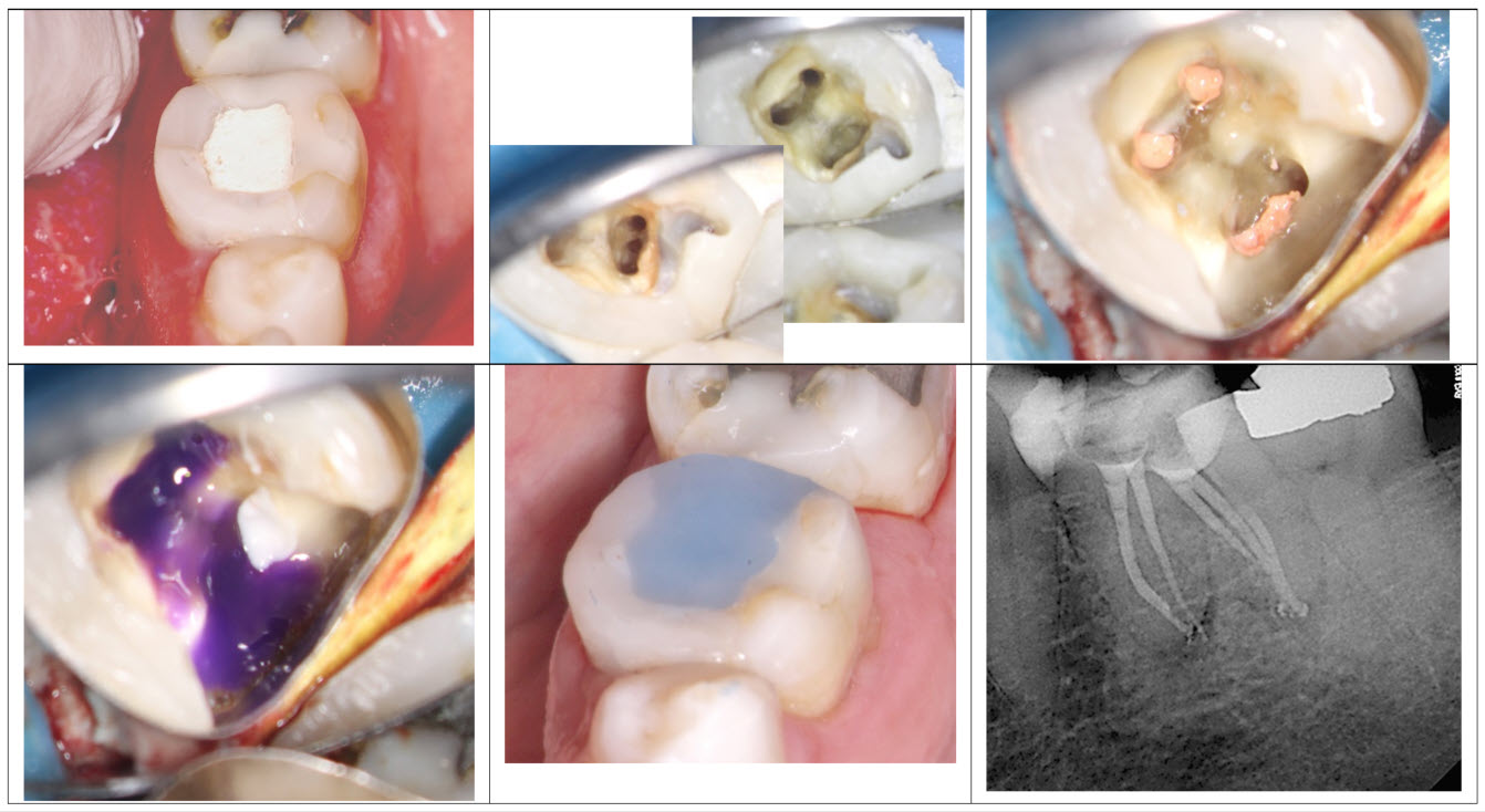
Once access was made we noted that the distal canal had three separate orifices with a common foramen. The canals were cleaned shaped and then packed and an off angle film clearly showed that they were separate and joined at the apex. This 3 canal system in the Distal root is quite unusual, since the “middle canal” is most often associated with the mesial root of the molar. The final radiograph does not show this complex anatomy because of the superimposition of the canals over each other that IS visible in the shifted . The case was closed with an orifice bond of Perma flow purple. The referring dentist had asked that I prepare a post space in the distal root so I selected what I felt was the best candidate for post space, prepared it and then filled it with calcium hydroxide prior to closing the tooth with a temporary. from this case we learned that the variability of anatomy in mandibular molars is infinite.

When we are exploring canal anatomy and mandibular molars we must always consider the possibility of a middle mesial or a middle distal canal that also requires treatment. Fortunately in most cases it joins to one of the other canals but in rare situations, it can have an entirely separate orifice and foramen. Failure to recognize this can result in persistent symptoms or ultimate failure of the case.
