BWs Crucial for Diagnosis
Proper Radiographic examination is crucial for good diagnostic technique. Periapical radiography in the mandible is sometimes difficult to achieve because of lack of patient cooperation, mandibular Tori or a small mouth. For that reason, periapical radiographs can sometimes be foreshortened and/or misleading. Bitewing radiography is important to be able to assess the Restorative margins for recurrent decay or deficiencies.
This patient was referred to me for Endodontic consideration of a mandibular second molar (#37). The tooth had been crowned with a full gold Crown and was quite mesially tilted. It was symptomatic and appeared to have a radiolucent apical finding of endodontic origin at the apex. The first molar had been extracted resulting in the heavy mesial tilt. The adjacent #38 was unopposed.
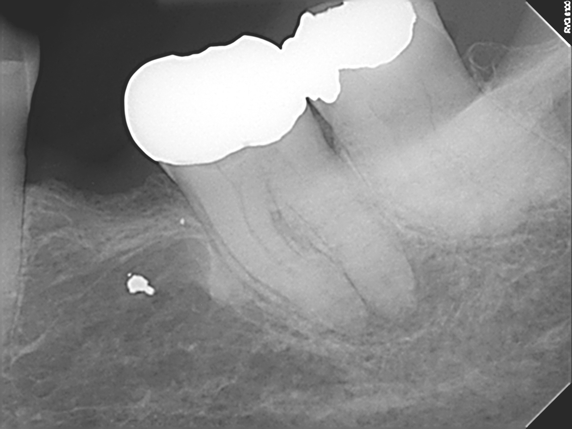
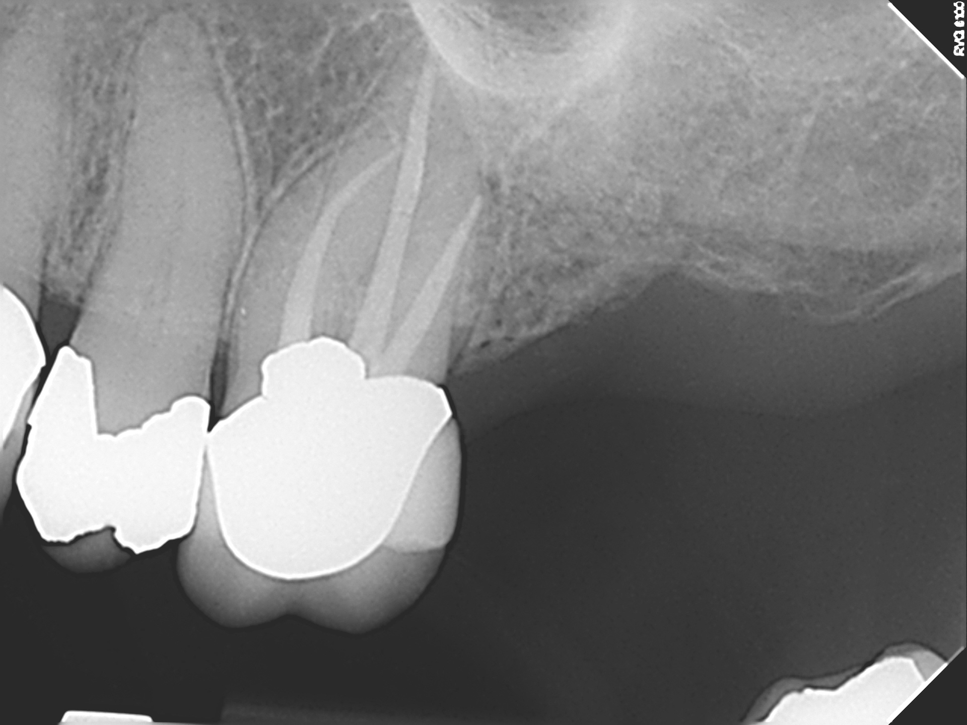
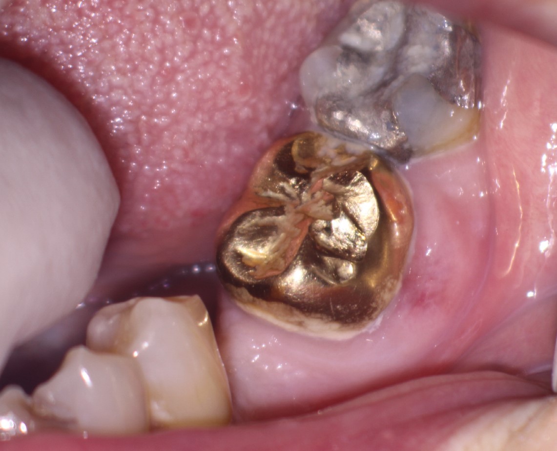
Outwardly, the tooth appeared to be relatively normal and there was no evidence of any problems with a cursory intraoral view, though a small amount of gingival discoloration was noted in the buccal gingiva. However, we know that more detailed examination can reveal significant decay underneath Crown margins. In this case a new PA and a bitewing radiograph revealed extensive buccal decay that was threatening restorability. I also noted that the patient’s hygiene was poor.
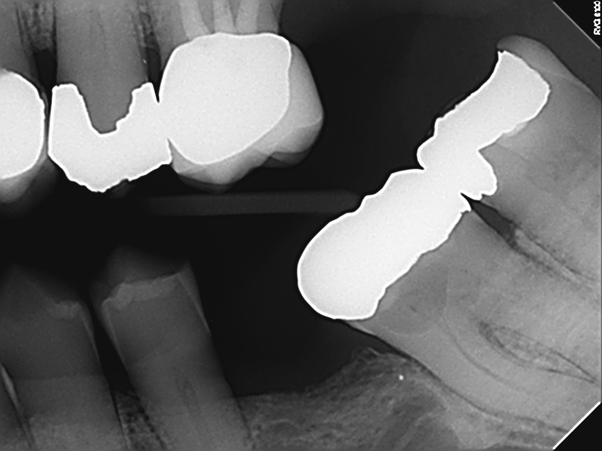
We had gone from a situation where the tooth may have just required Endodontic treatment through a crown, to one where it is very likely that Endodontic treatment, Periodontal surgical procedures (crown lengthening or graft?) and then new full Crown restoration would be required. The decay was at the osseous crest. Adding up the cost of these procedures, we began to approach the cost of an implant. Implant placement in the position of #36 may, in fact, offer a better option because the implant in the position of #36 will be in a more favorable occlusal relationship with the opposing Endo treated, crowned maxillary first molar, #26..
Had a proper bitewing been taken and examined prior to the referral, it is likely that the patient would not have been referred for treatment or examination because the level of the decay would have been ascertained prior to referral. Or, if the patient wanted to try to keep the tooth, the referring Dentist should have immediately removed the crown, excavated the decay and satisfied himself that the tooth (and all procedures necessary to restore it) were acceptable to the patient prior to referral for Endodontic treatment.
The angulation of images taken in the mouth is extremely important. One of the biggest problems that Endodontists see with images taken by referring dentist is poor angulation and inability to image the tooth with sufficient diagnostic quality. In most cases this can be easily remedied with the conscious use of paralleling devices whenever any radiograph is taken.
There will always be situations in which patients are either non compliant or the standard parallelling instrument may be difficult to use (Uncooperative patients, tori, gagging, small mouths, etc). In those cases , experience and intuition are necessary along with several different views to be able to image all structures necessary to properly evaluate the tooth and decide whether the tooth merits further treatment or extraction. cbCT imaging can also be helpful though it has its limitations in mouths restored with multiple adjacent implants, alloys, posts and Endodontics.
