Management of Dens Evaginatus
This 17 year old Asian female patient was sent to me for consideration of a Virgin mandibular second molar on the right side. The patient had a history of pain in this tooth but was not sore on presentation. the patient had been placed on antibiotic medication for the previous six days.
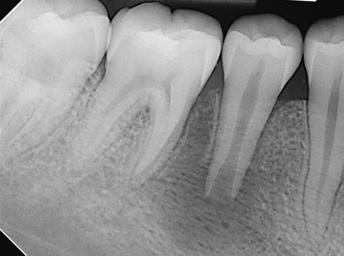
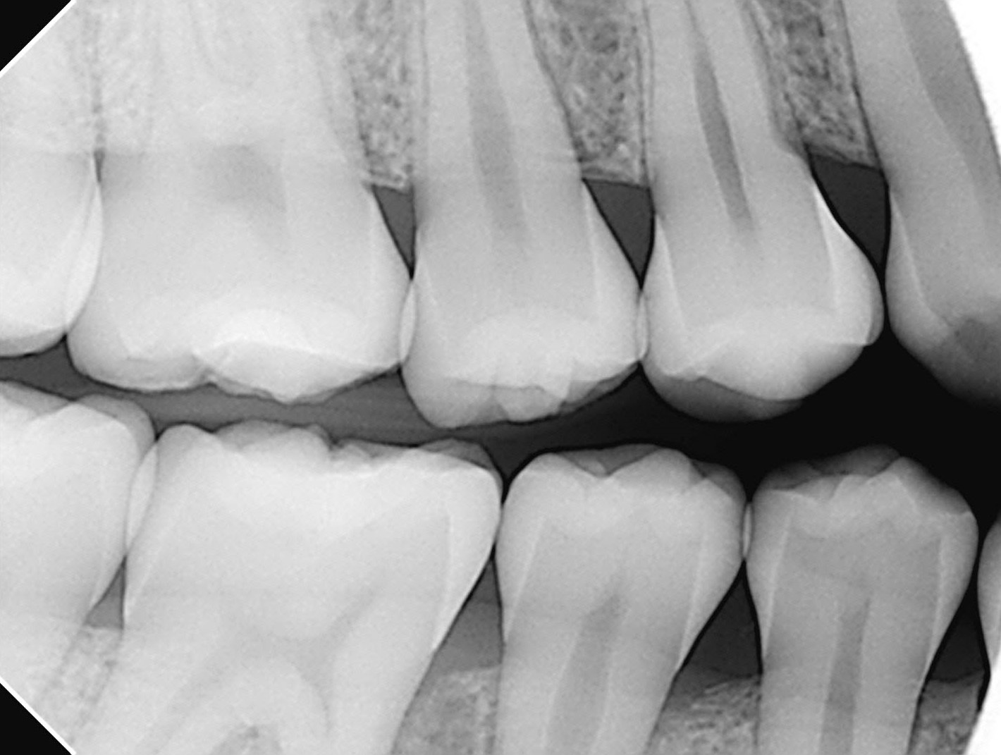
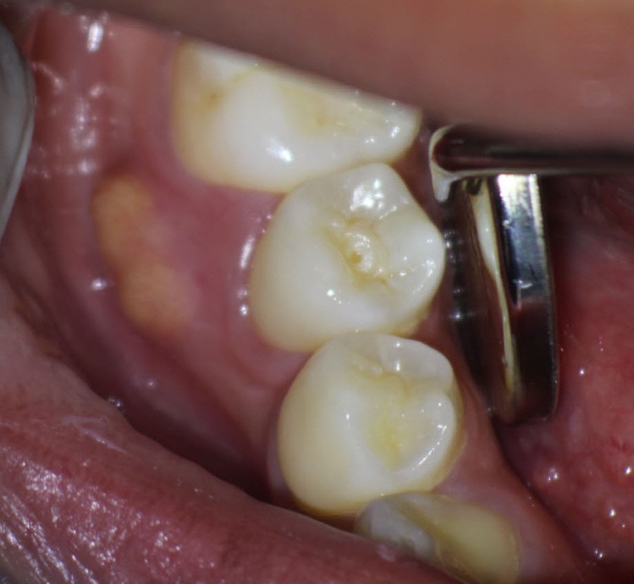
Radiographic examination showed classic Dens Evaginatus. Apical development was incomplete and there was a diffuse radiolucent area associated with a blunderbuss apex . There was an associated draining buccal sinus and the gingiva was slightly swollen and discolored. Palpation of this area was minimally positive. All other findings were normal. Periodontal probings were within the range of normal.Pulp tests were non-responsive. A Diagnosis of Dens Evaginatus, incomplete apical development and chronic periapical periodontitis was made.
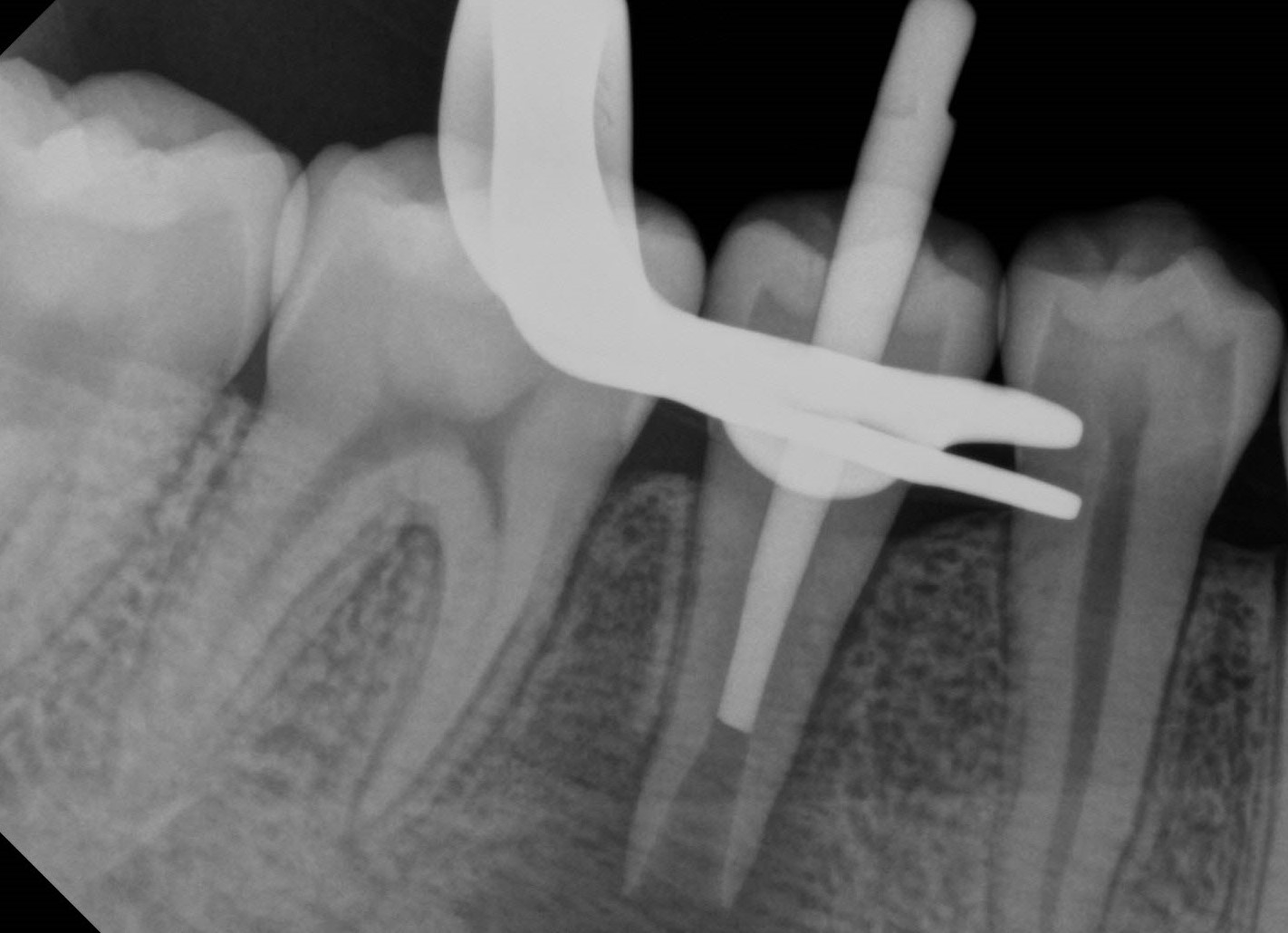
A rubber dam was placed in the tooth was accessed without local anesthetic. The access was rinsed with NaOCl 5.25% and attempts were made to ascertain the level of any vital tissue in the canal. Paper points were used to test the area and established the rough estimate as to where the necrotic pulp ended and the apical tissue started. I used a custom rolled gutta Percha cone to radiographically check where this level terminated. Calcium Hydroxide medication was inserted into the canal and gently packed with large paper points and Schilder Pluggers. The patient was instructed to return at three month intervals to assess symptoms and the periapical area. The sinus closed and the apical radiolucency began to resolve.
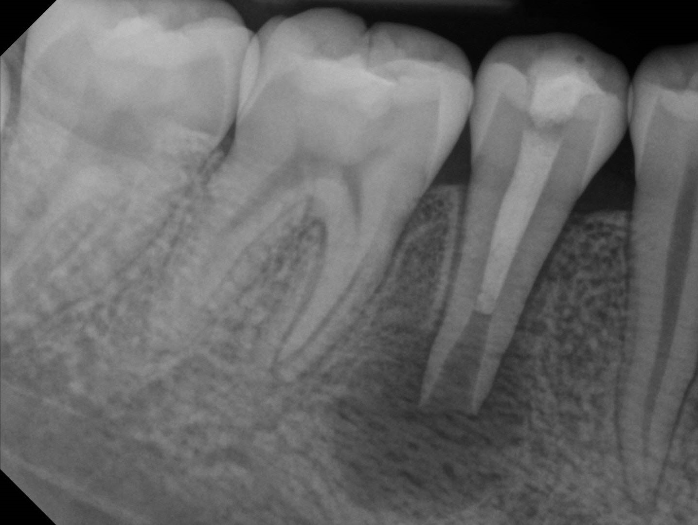
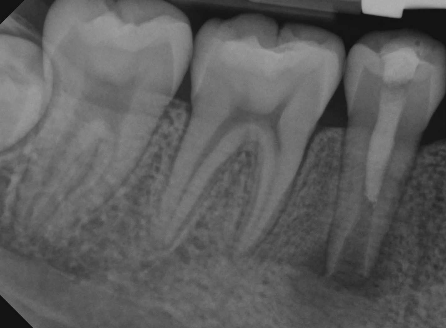
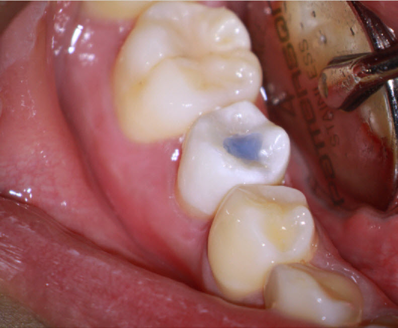
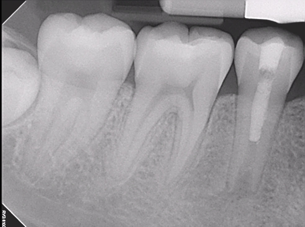
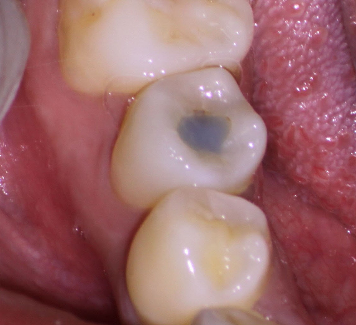
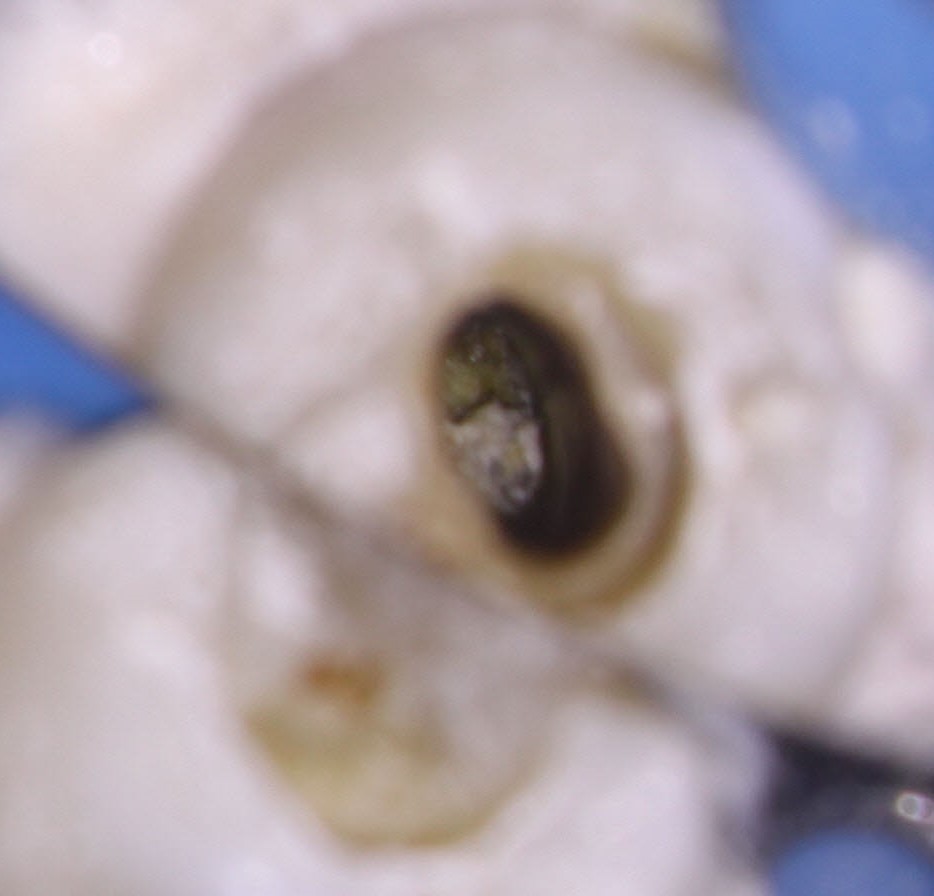
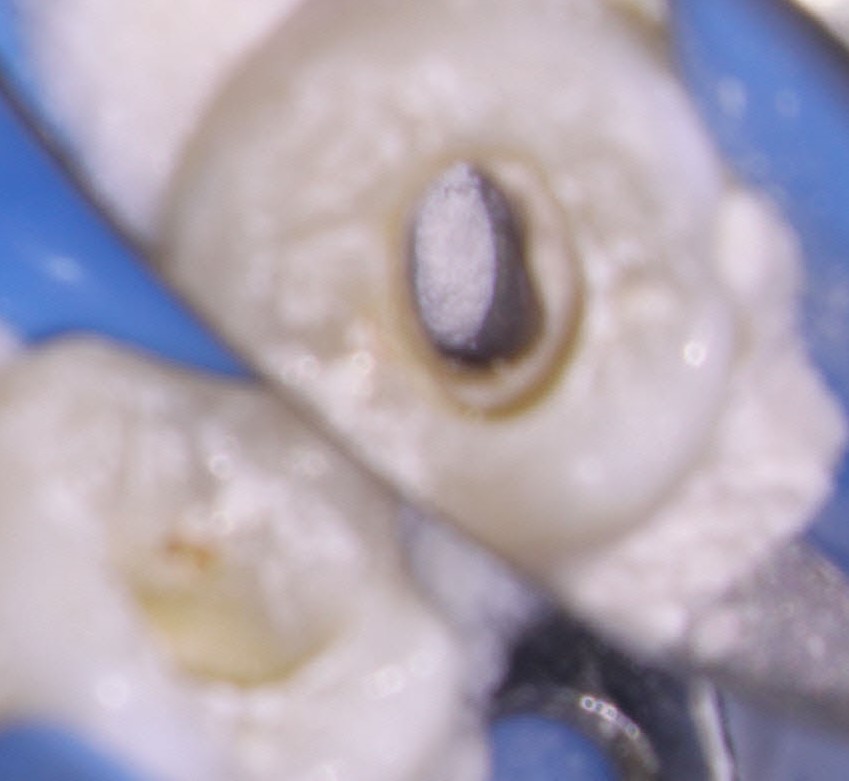
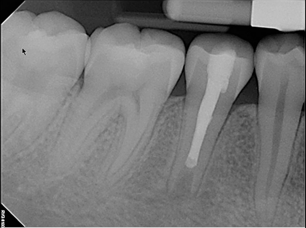
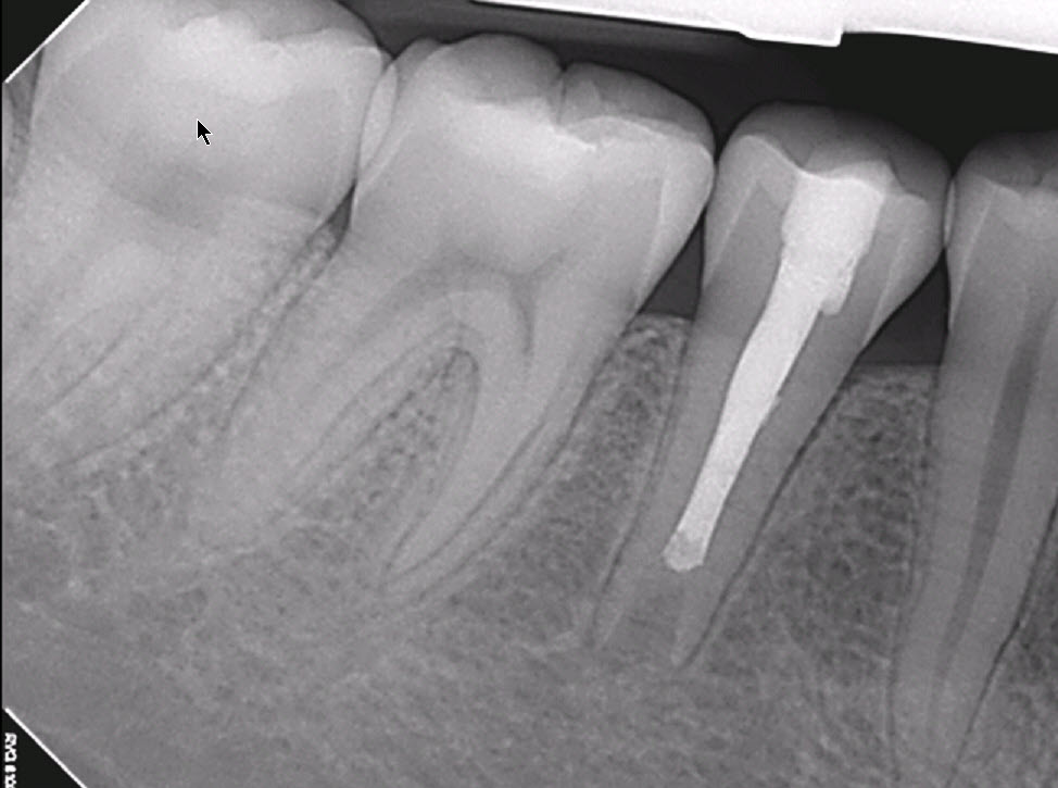
Approximately 6 months later, the area has resolved sufficiently to allow me to obturate the canal and the patient was totally comfortable . Obturation was completed by filling the apical section with MTA, followed by gutta percha filling in the coronal part of the canal. A composite and fillingwas used to close off the access.
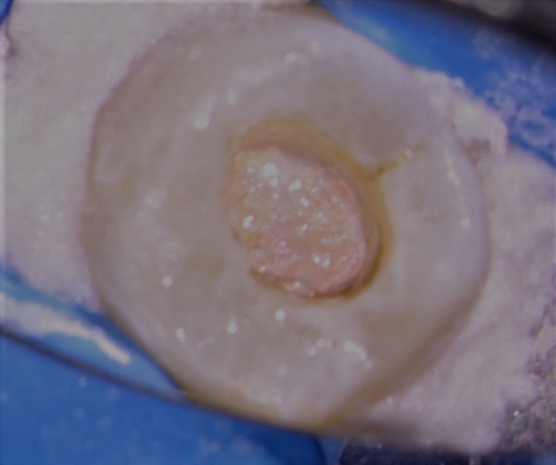
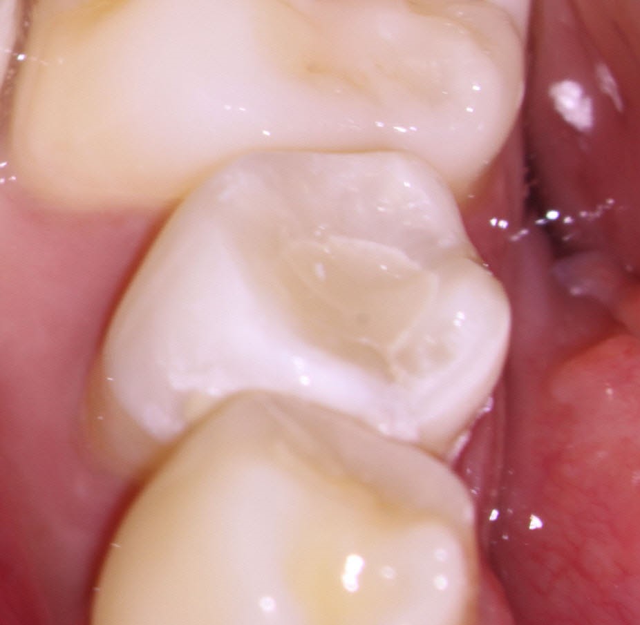
The most critical part of managing the case is to determine the level at which we wished to terminate the cleaning and shaping procedures and being able to control the obturation materials to the point where no excess is extruded. No barrier techniques were used in this case. This is a slow process and patients have to understand the slow pace at which the pathology resolves
