Iatrogenic Oral Surgery Fixation Complicates Case
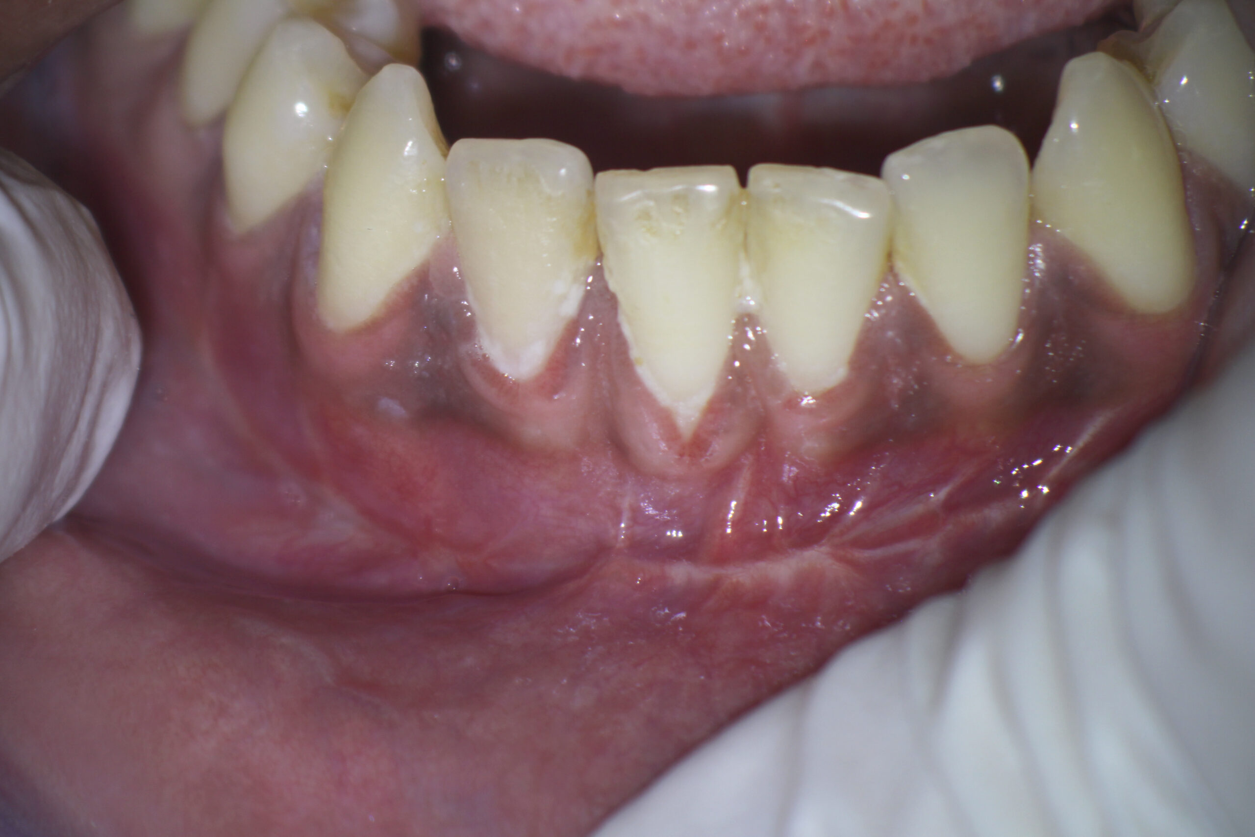
This 27 year old male patient was referred to me after having had a traumatic incident which resulted in the fracture of his right mandible . The patient had been seen by an Oral Surgeon for repair, which involved surgical fixation of the segments with screws and bars. Patient was having continuing symptoms which are described a

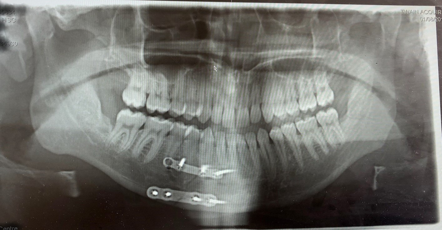
I asked the referring dentist to contact the Oral Surgeon and send any pertinent information he felt was important. I received a photo of a panoramic image that apparently was taken with an iPhone by the referring Dentist.

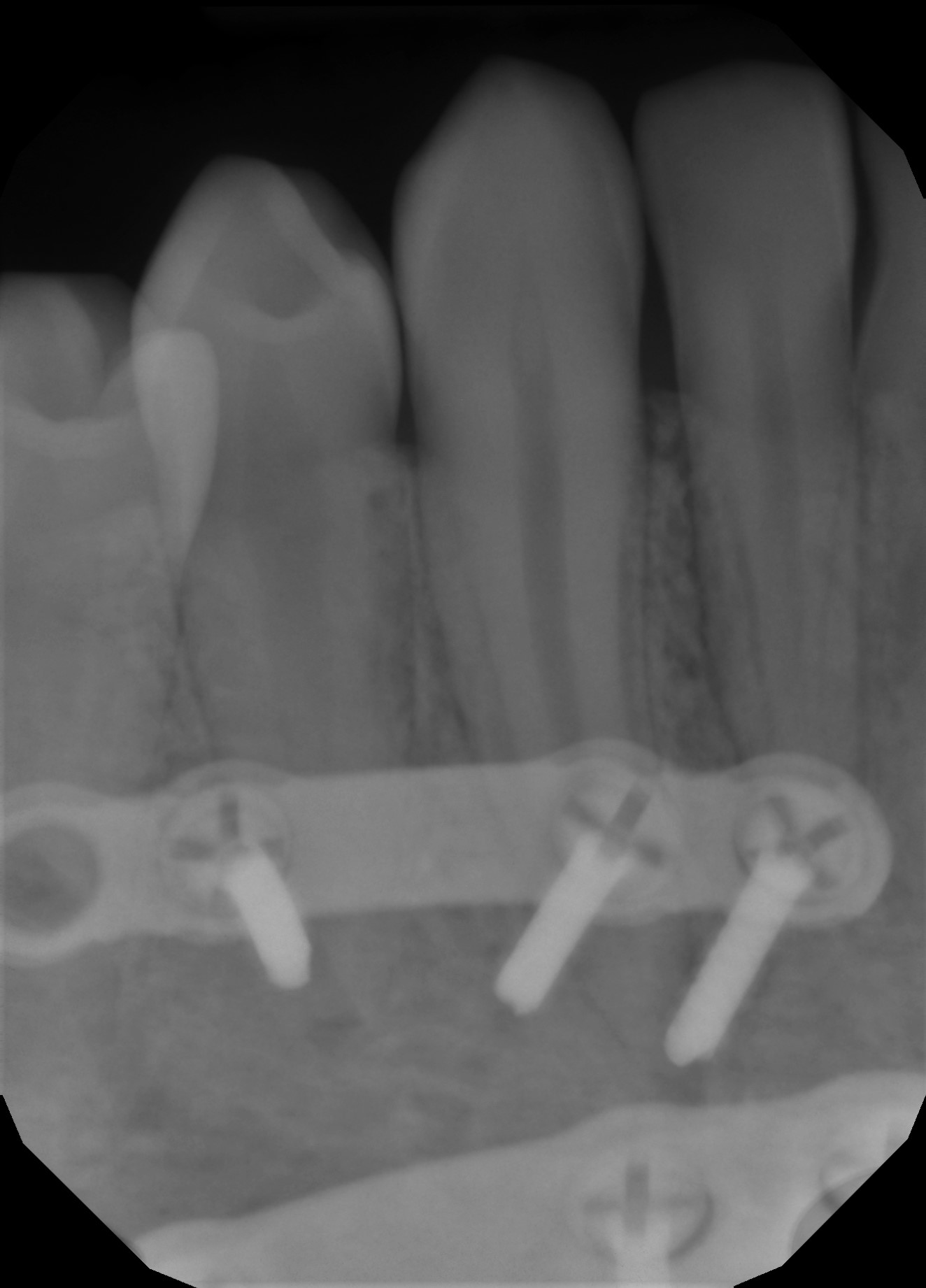
cbCT examination of the area showed an oblique fracture of the mandible that had been repaired with fixation. Unfortunately the oral surgeon placed the screws of the upper level fixation directly into the roots of the canine and second premolar. Apparently, he realized he realized the error in the second premolar and the screw was removed, leaving a void and the root as visible on the cone beam tomograph. The cuspid screw had been placed right through the cuspid root and out the lingual plate!!! The case was certainly unusual and something I had never seen before.


I explained to the referring dentist that our options were quite limited. Both the cuspid and the premolar would require Endodontic treatment due to the iatrogenic perforation of the roots by the screws.
- We could attempt Endo treatment of tooth #45 using conventional methods but the perforation would essentially make the case an open apex situation. Because there was no pulpal pathology prior to the iatrogenic perforation, (I assumed that these teeth were virgin and pulps vital) it may be possible to place MTA to the level of the screw hole and then simply fill the rest of the canal and access. We would have to determine how well the area healed once the Endodontic treatment had been completed on this case.
- However, in order to treat the problem, the fixation would have to be removed so the screw could be removed from the apex of the cuspid. Once the fixation hardware was removed, we could visualize the apices more clearly with radiographs. We would then treat the cuspid similarly to the premolar, attempting an open apex type of treatment with MTA as necessary.
I may have considered performing Endodontic surgical procedures on these teeth myself but I did not want to take the responsibility of removing the screws and plate because I did not place them originally. We also did not want to have the patient undergo separate surgical procedures for (1) hardware removal and (2) Endo surgery, if necessary.
If subsequent Endo surgery was required , it could be performed on the damaged apical segments without interference from the hardware. This would necessitate a second surgical procedure, which is something I’m sure the patient wanted to avoid, considering what he had already endured, dealing with his fractured mandible. Also, with each subsequent surgical procedure we run the risk of paraesthesia when working in the area close to the mental foramen.
In the era of digital imaging, this kind of iatrogenic error is surprising and totally unnecessary. Prior to the surgical procedure the surgeon should have examined the anatomical position of the roots of these teeth, scanned the case, and calculated screw positioning to avoid any possible iatrogenic damage. Surgical stents could have been fabricated, ensuring precise, accurate proper screw position and depth.
I suggested that the patient return to the oral surgeon to have the hardware removed. Once there was evidence of healing of the fracture we would attempt endodontic treatment. As the patient did not return for follow up, I have no knowledge of how this case eventually was resolved.
