Retrograde Filling of Unfilled Canal
A 59 year old male patient in good health was seen for endodontic consideration of tooth # 22. He presented with a 3-unit bridge #11- #22 and had a history of a draining buccal sinus adjacent to pontic #21. He was otherwise asymptomatic. The patient was quite satisfied with the clinical appearance of the bridge, so the both aesthetic and financial reasons, he preferred not to have the bridge remade, if possible.
Percussion, palpation, chewing and periodontal tests were WNL.

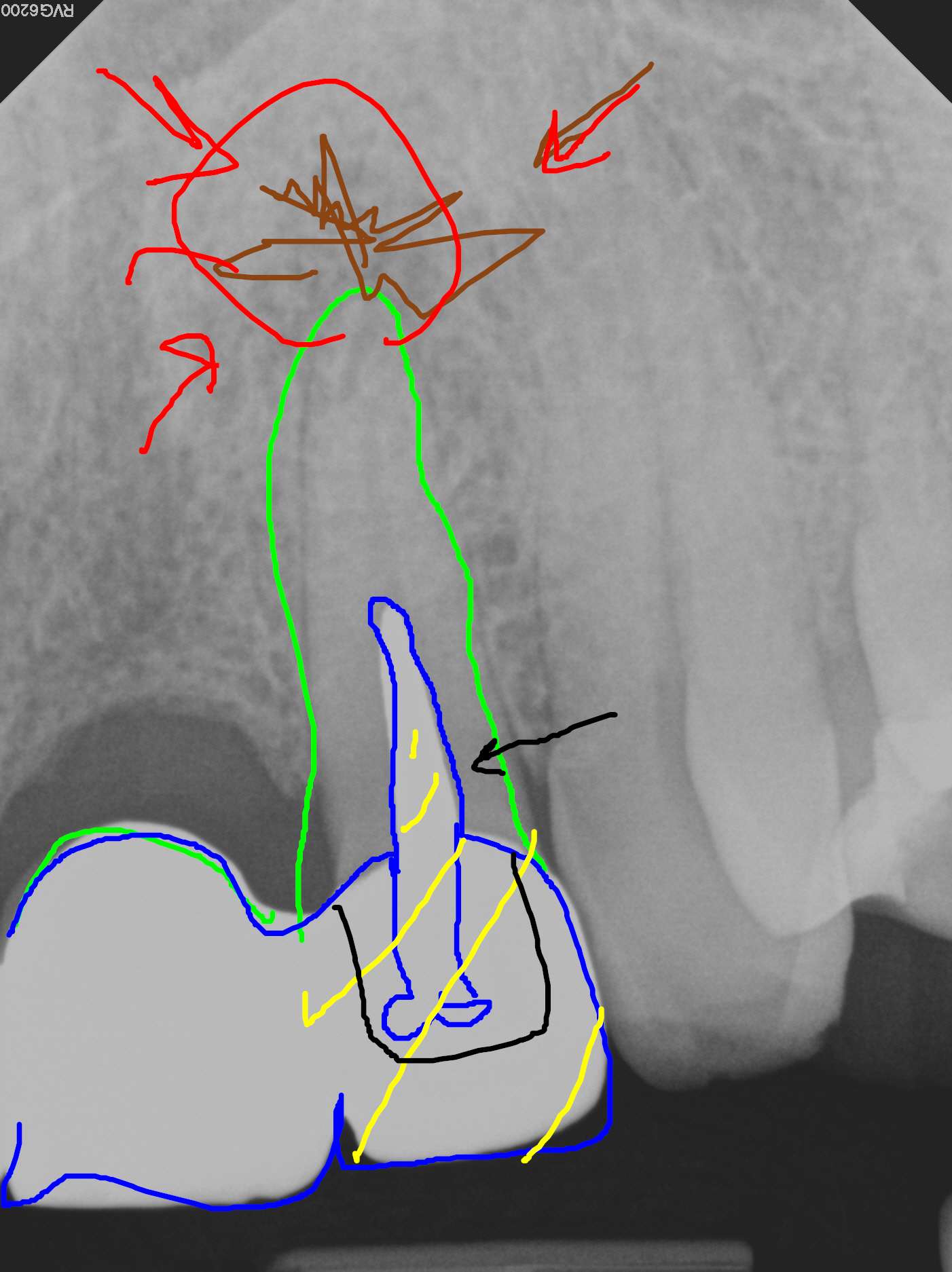
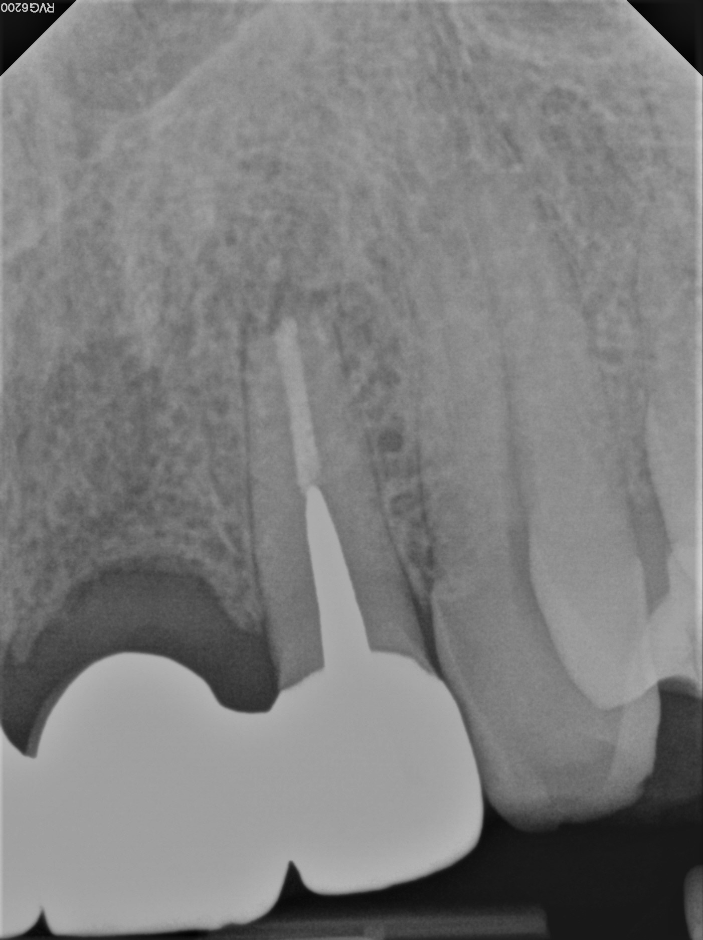
Radiographic examination revealed a cast post that appeared to have been placed in a root without previous endodontic treatment. A persistent periapical radiolucent finding was noted at the apex of #22. A diagnosis of Chronic Periapical Periodontitis was made secondary to the unfilled necrotic canal.
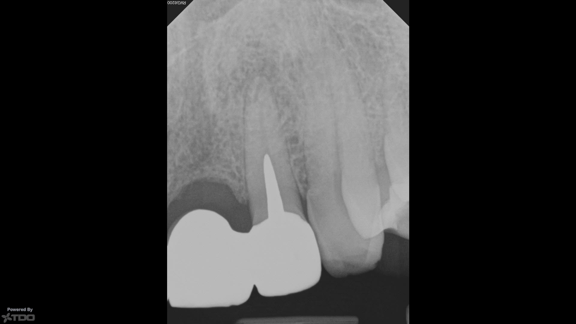

There was a hint of dark discoloration, seen through the palatal aspect of the porcelain crown, which suggested the underlying post was likely cast metal. This was consistent with the conical shape of the post and radio-opacity as seen on the radiograph.
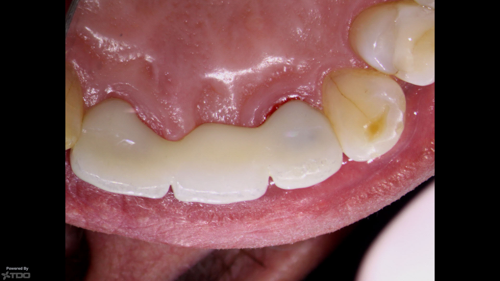
Because the patient was happy with the clinical appearance of the all ceramic bridge, bridge removal was deemed to risky. I was unsure whether the cast post in #22 was separate or whether the crown/post was one unit. Attempting to remove it through the crown (to retreat the case conventionally) would likely be impossible, or result in loss of retention of the abutment. In any case, bridge removal was ruled out.
After reviewing alternatives, we chose surgical endodontic retreatment consisting of root resection and retrograde filling of the unfilled canal.
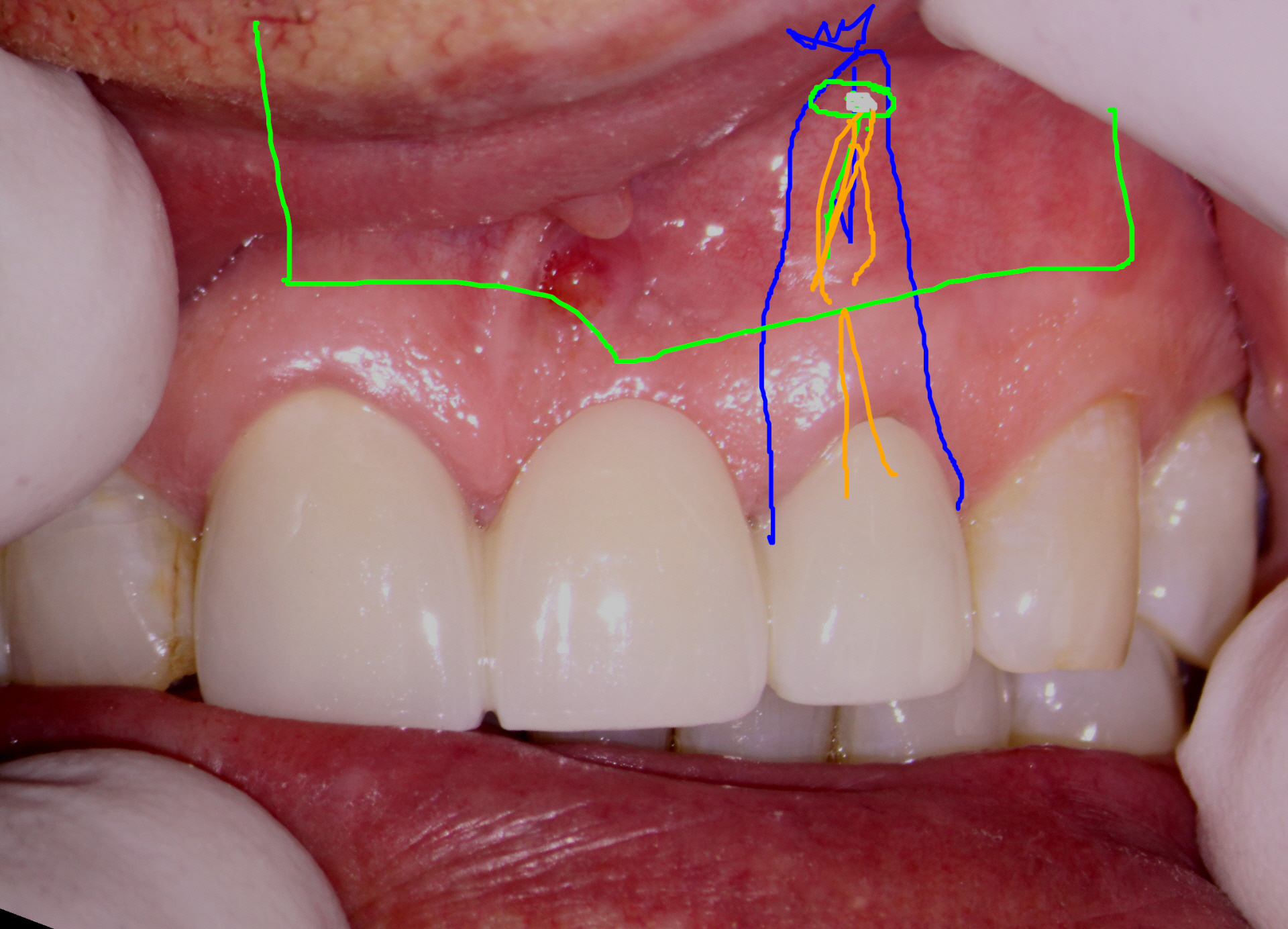
The patient was anesthetized and a BU flap (Luebke – Ochsenbein) was raised to expose the periapical area.
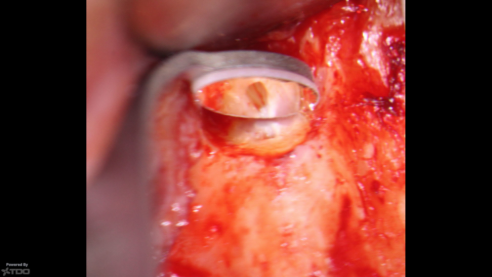
The root was resected minimally and a piezo file adapter was used in conjunction with a piezoelectric ultrasonic tip (Satelec P5) in order to prepare the length of the canal in a reverse manner. The canal was irrigated with solutions of NaOCl 5.5% , acidified NaOCl and 2% CHX with close suction. The canal was dried with Ultradent tips, Stropko Syringe and paper points.
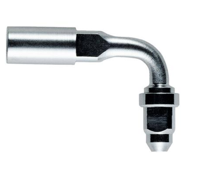
90 Degree Piezo Endo File Adapter
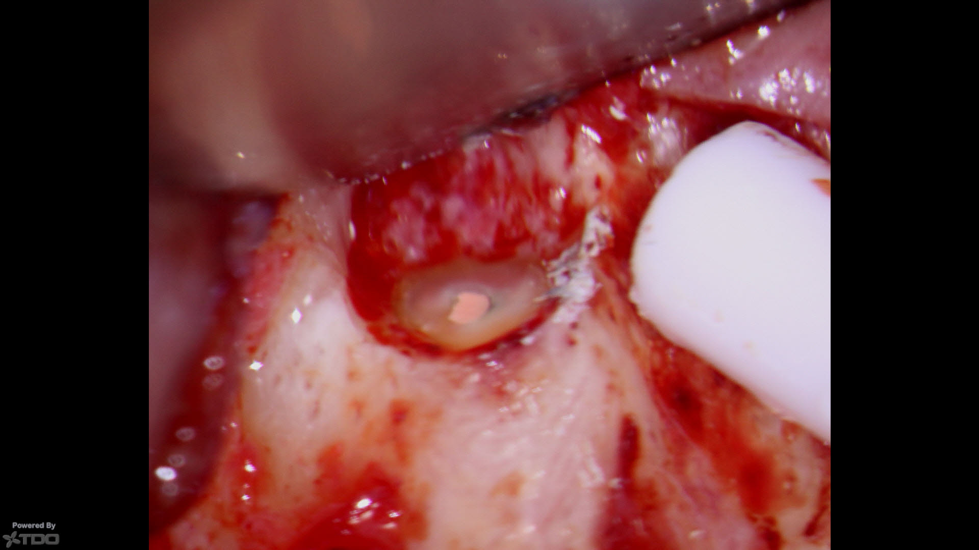
The canal was filled with Kerr sealer (applied with a paper point) and injectable Gutta Percha (Hot Shot) with a tip bent at 90 degrees to allow for deep access into the canal. Retrograde pluggers were used for condensation and the gutta percha was burnished with an Egg burnisher. No other retrofilling was deemed necessary as I felt seal was adequate without further retropreparation and loss of root dentin.
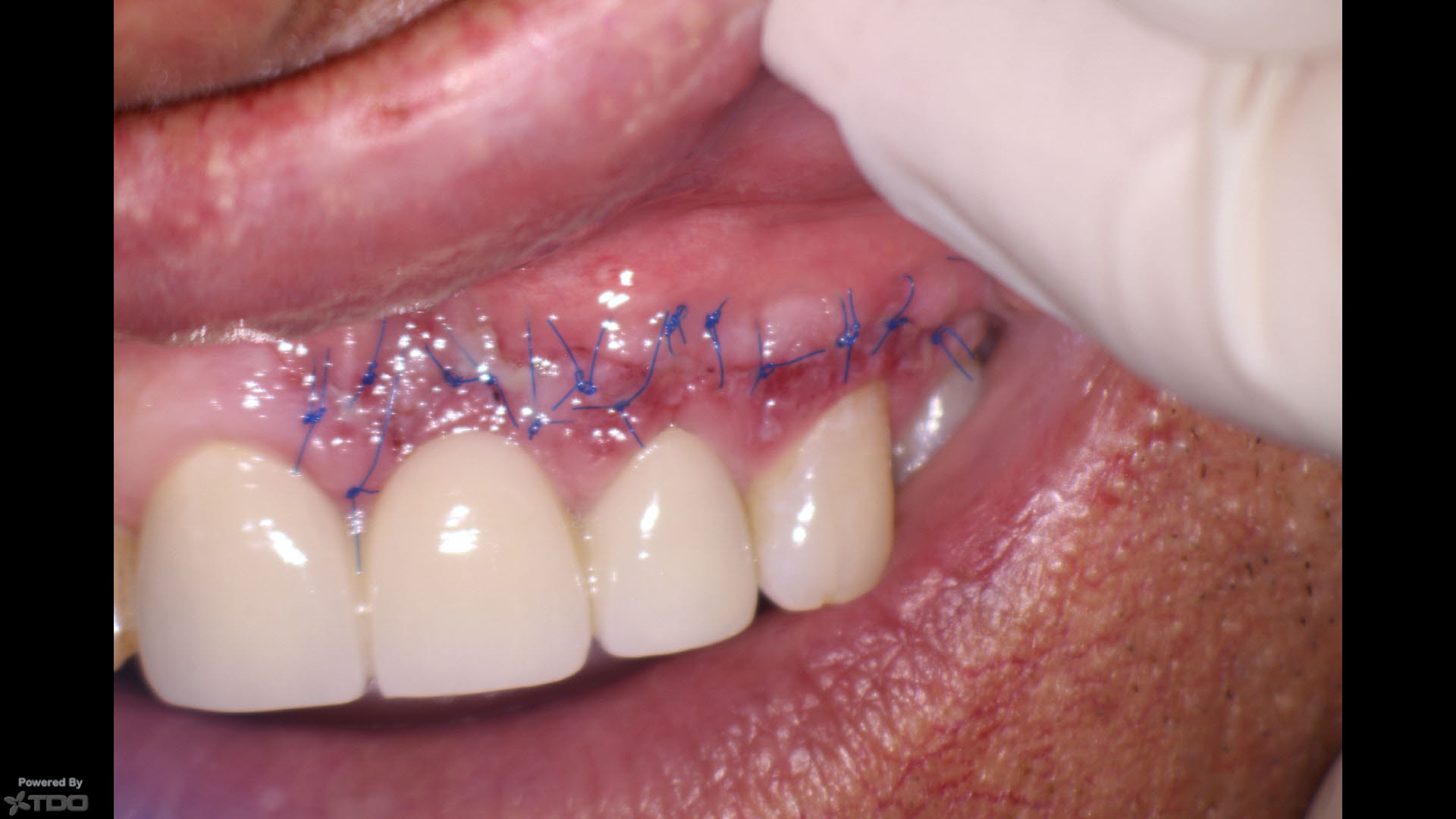
The flap was sutured with 6/0 Proline interrupted sutures.
The flap was sutured with 6/0 Proline sutures.
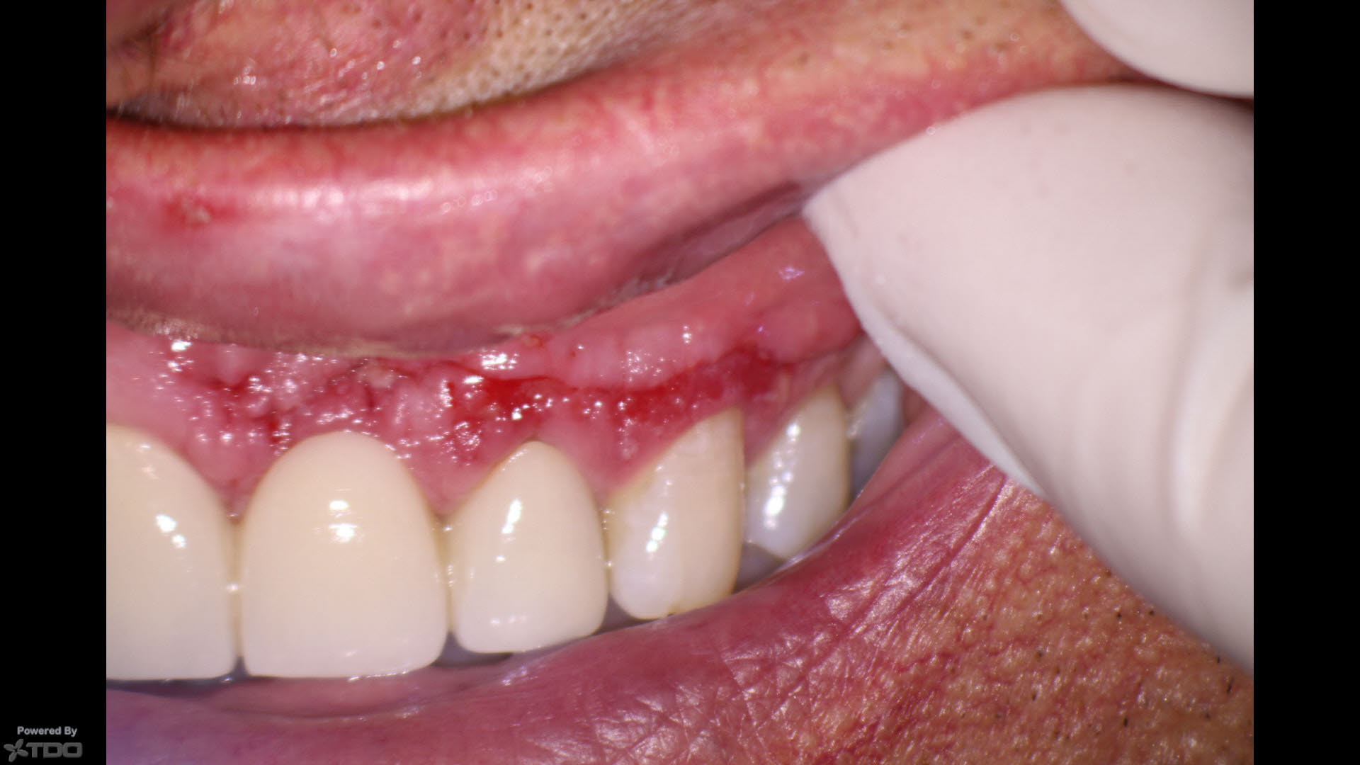
The sutures were removed 3 days later and the area and the flap are healing without incident.
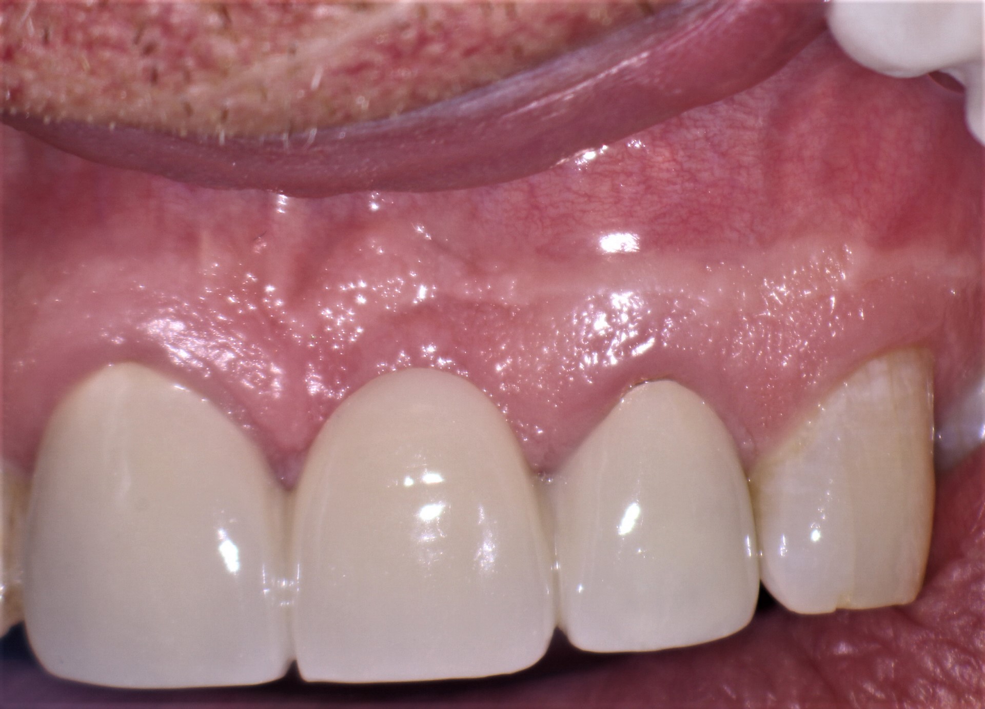
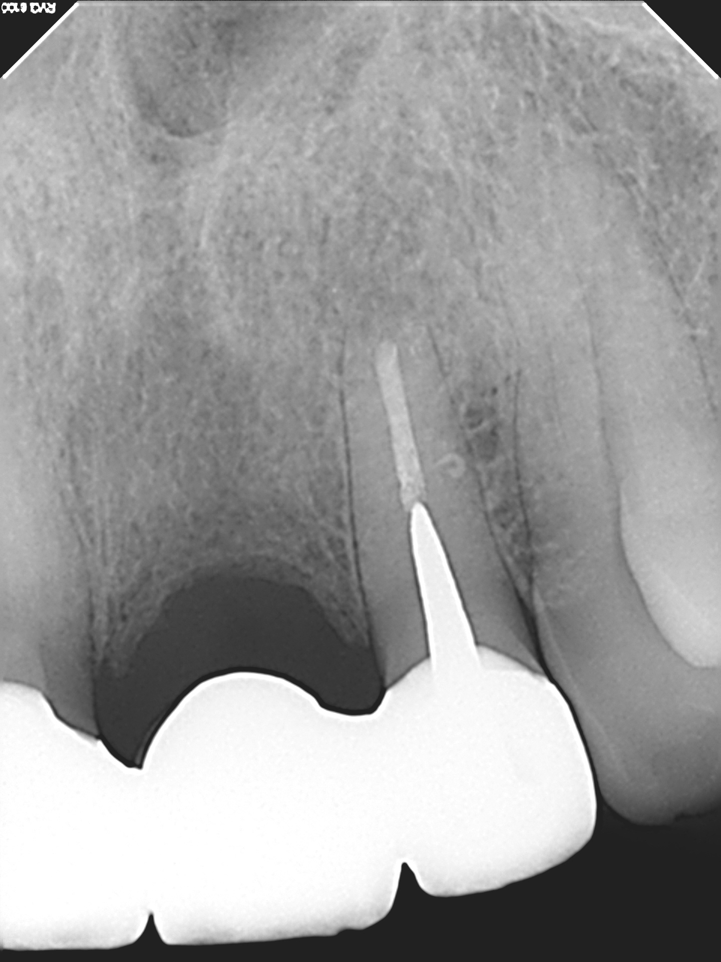
3 Mo. Soft tissue check shows good healing, normal periapical bone and asymptomatic patient.
