A 3 Rooted Cracked Virgin Maxillary Premolar
A 33 year old male patient was seen referred to me for examination regarding symptoms arising from his right maxillary 2nd premolar. The patient’s current complaint was discomfort when chewing down on this tooth. The patient had recently had an implant placed in the position of the first premolar .
The patient was awakened at night with the pain. The tooth was not sensitive to hot or cold stimulus.The tooth hurts only if he bit on it. The Patient has been placed on Antibiotics for previous 3 days by the referring Dentist (!!??). The patient has taken pain medication(s) at various times.
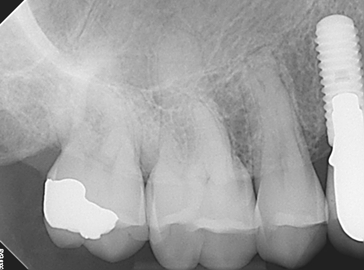
Figure 1 : Preop PA
Pa suggests multiple rooted 2nd premolar
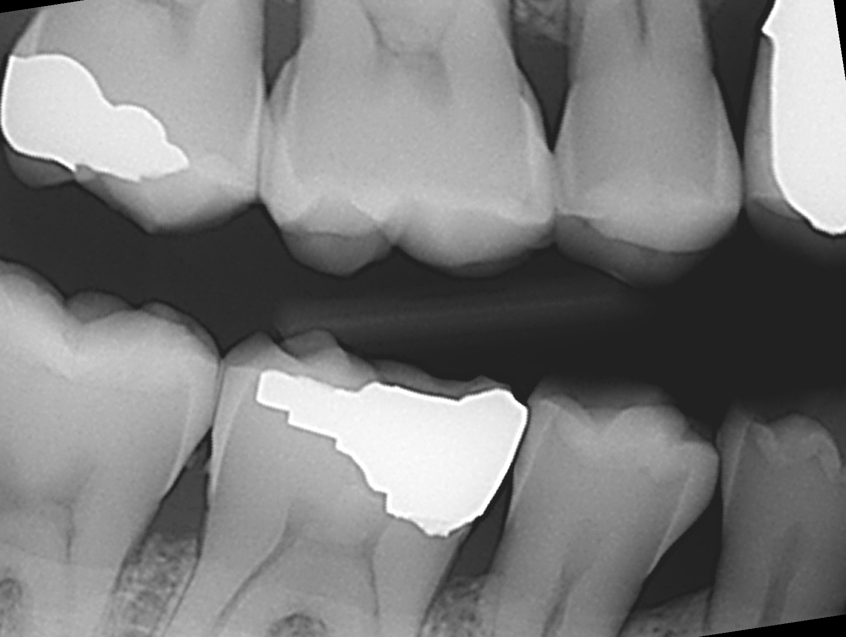
Figure2 : BW radiograph
No restorations visible
Clinical appearance was that of a virgin tooth. This second premolar showed positive responses to percussion and chewing. Cold and hot tests were non-responsive. Perio probings were WNL. Transillumination appeared to be positive for cracked tooth through the central developmental groove. No abnormal facets were noted and there was little evidence of bruxism.
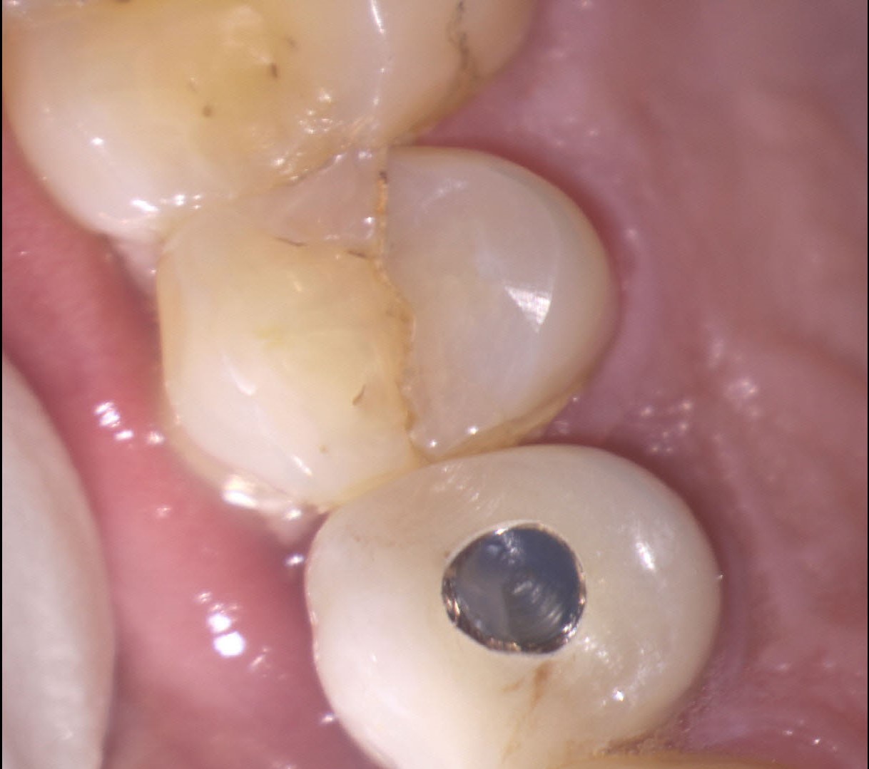
Figure3 : Preop image shows stained M-D crack
Transillumination also was positive for M-D crack
Conventional radiography showed no abnormal periapical radiolucent findings but the appearance of the roots suggested it may be a 3 rooted premolar. cbCT imaging confirmed the presence of 2 buccal roots and one palatal root.
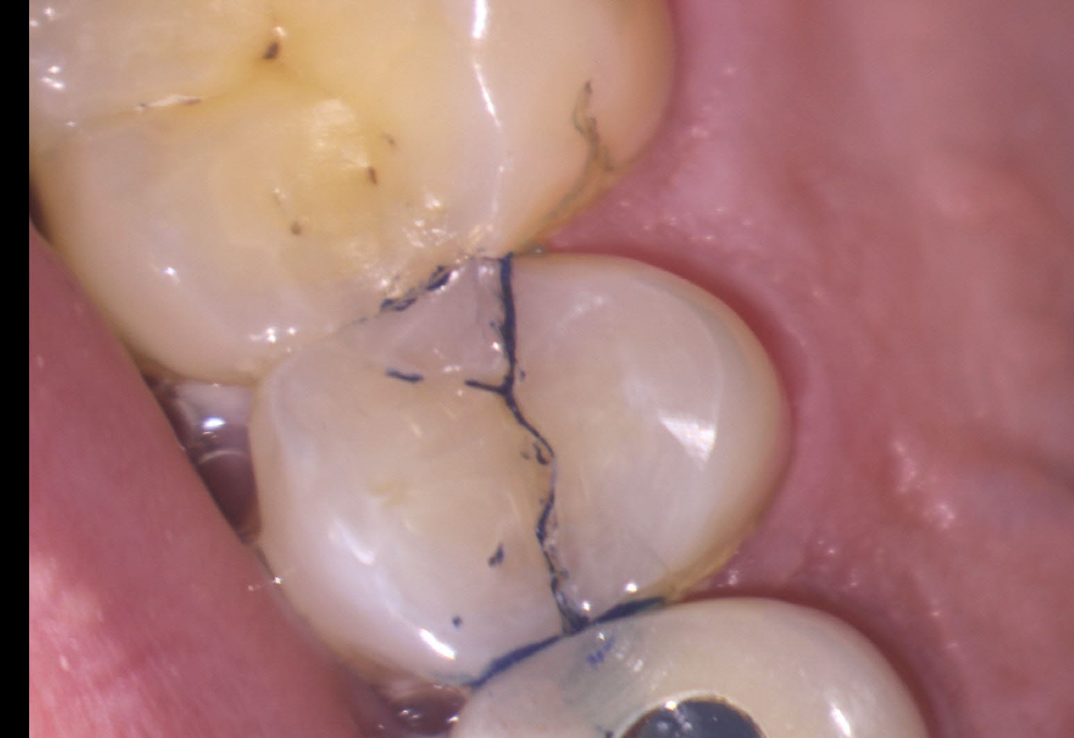
Figure 4: Methylene Blue Stain
Staining the crack can sometimes help explain the crack to patients.
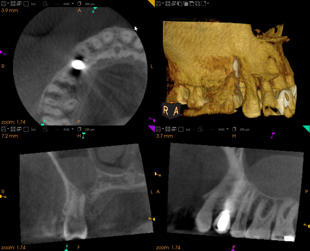
Figure 5 : cbCT imaging showing 3 roots
A diagnosis of acute periapical periodontitis secondary to pulpal necrosis (caused by a crack) was made and treatment options were explained to the patient. I would perform endodontic treatment on the tooth, relieve the occlusion and insist that the patient be seen by he referring dentist in the next 48 hours for preparation of the tooth for full crown restoration. Assessment of the depth of the crack (on the M and D sides) would be done with the aid of an SOM during treatment. The fact that periodontal probings were WNL was a promising sign but I cautioned the patient that the long term prognosis of the tooth was uncertain.
Note: In cases where the tooth CANNOT be restored immediately, provisions MUST be made to either temporize the tooth with a temporary crown or Cu/Ortho band.
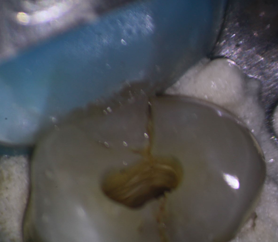
Figure 6: Access
A brown crack line is visible running down the D part of the crown. The extent of these proximal cracks and any associated Periodontal pocketing are crucial in establishing prognosis and determining whether the tooth merits continuation of treatment.
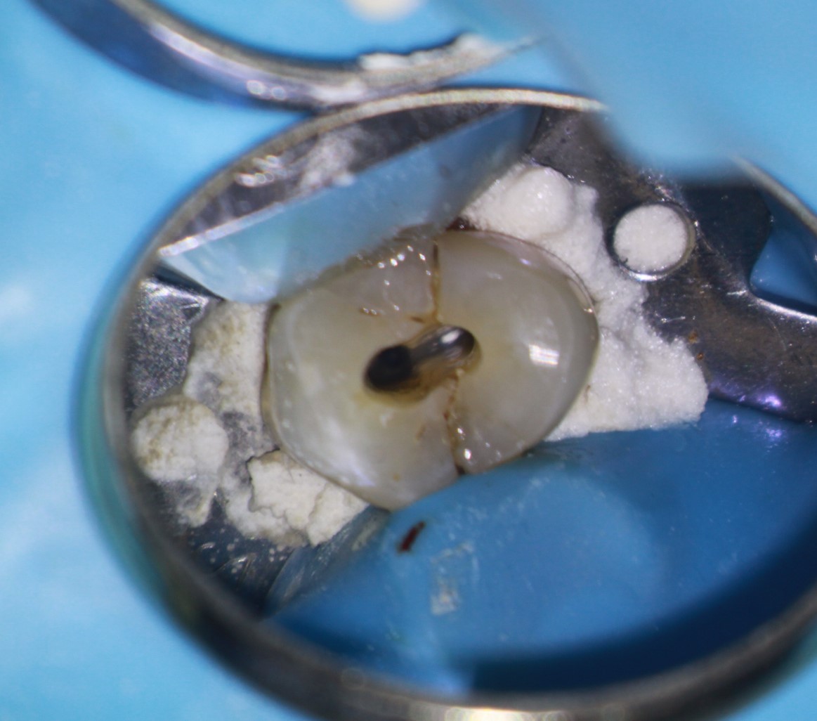
Figure 7 : Canals prepared
Conservative access preserves coronal tooth structure. Even with this level of magnification, identifying and treating the 2 buccal canals can be difficult. cbCT imaging is essential.
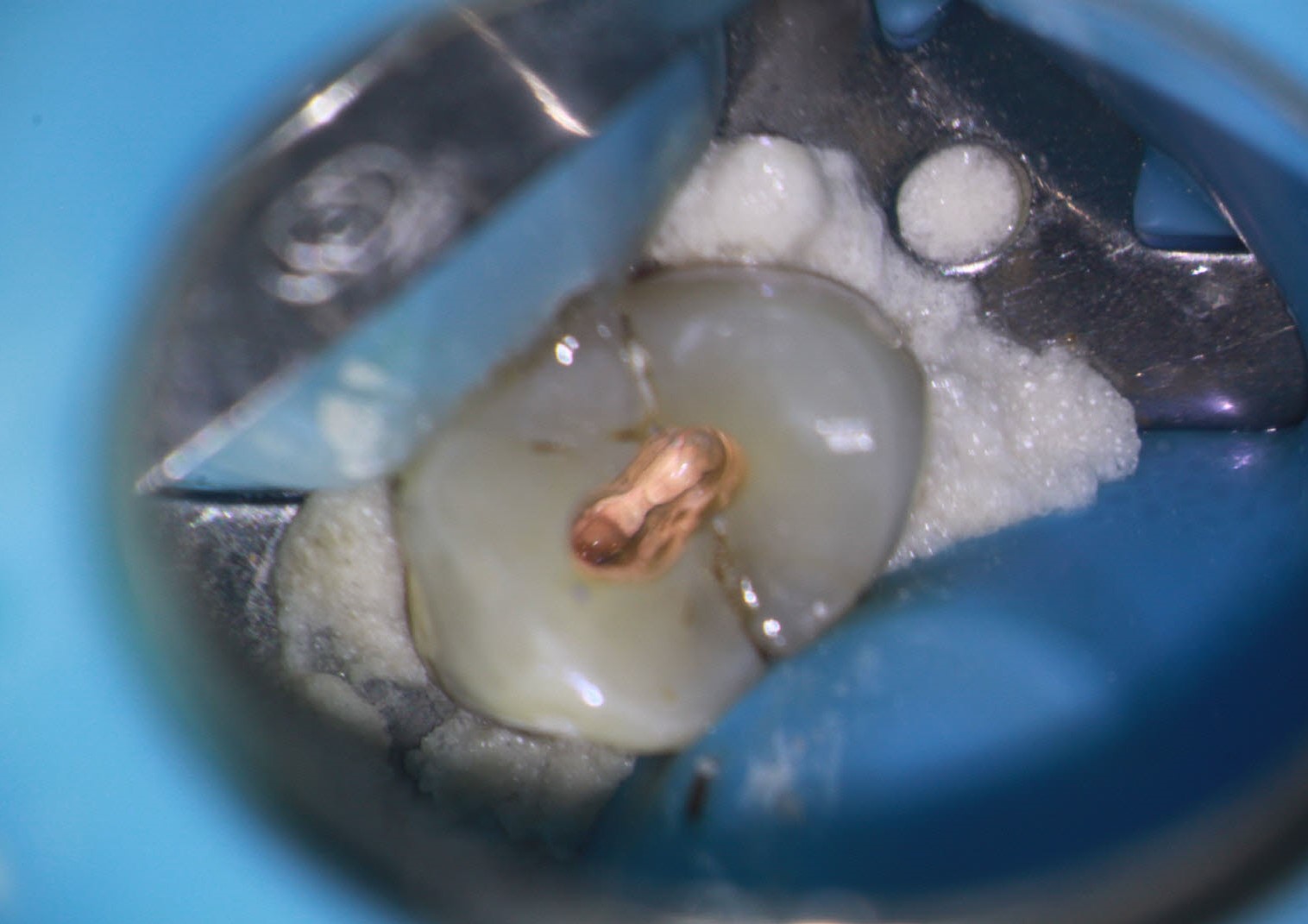
Figure 7: Obturation complete
Vertical compaction of warm gutta percha (cones) used for canal filling.
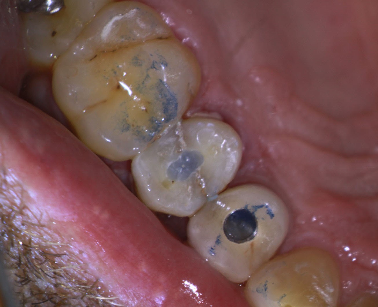
Figure 8: Access closed with Bonded CorePaste filling
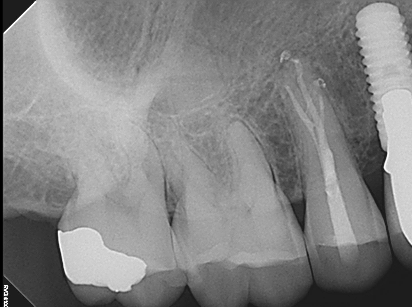
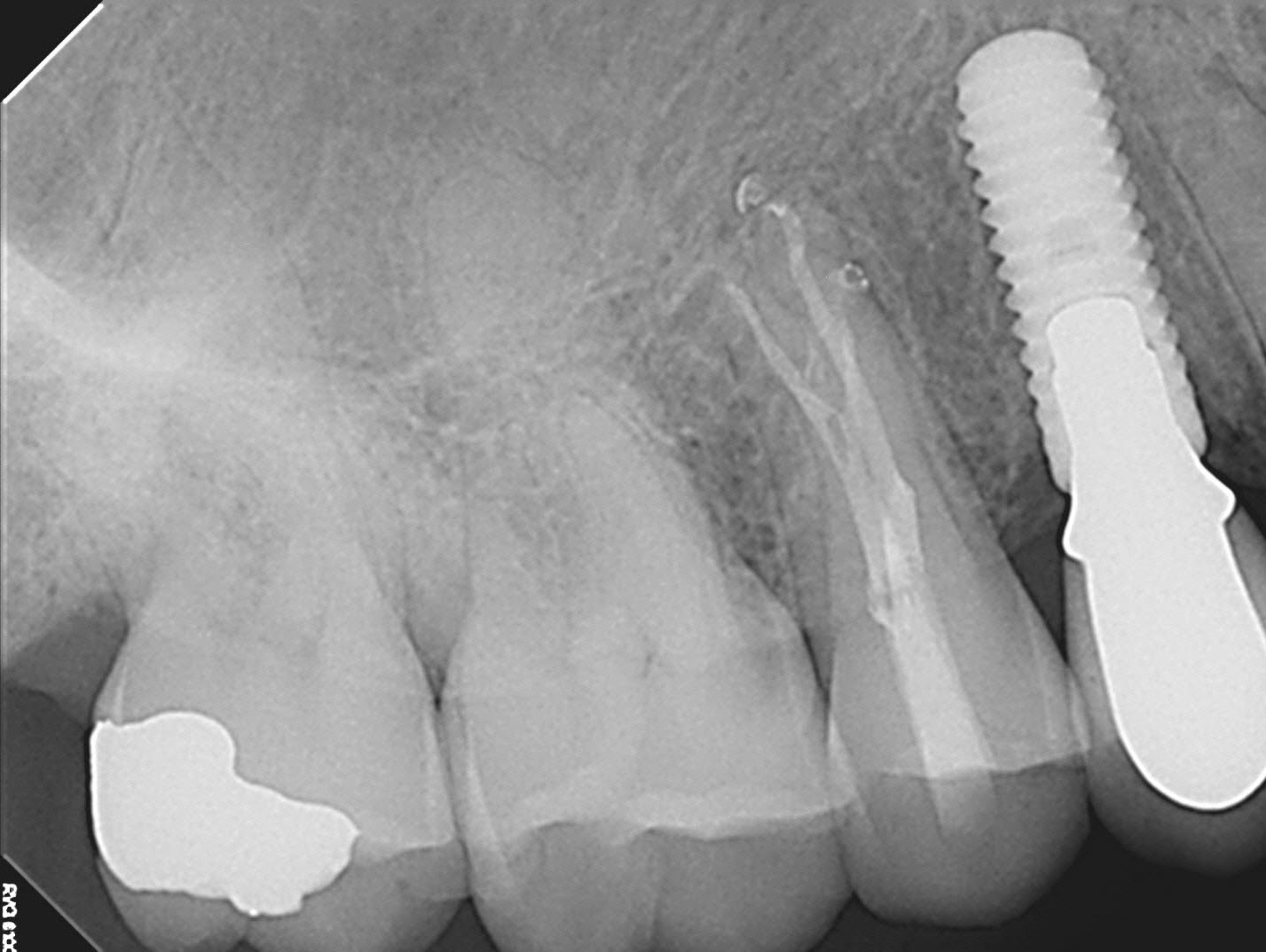
Treatment was completed with minimal access and the patient was referred for immediate cuspal protection restoration the following day.
Examination of the rest of the patient’s dentition did not reveal any abnormal faceting or heavy occlusion. It is possible that the initial crack may have started while the patient was waiting to have the adjacent implant restored. Or he simply may have had a bit of bad luck biting on a popcorn kernel, almond or other hard food.
cbCT imaging was invaluable in confirming the presence of three roots in this tooth and allowing us to preserve as much dentin as possible by not “hogging out” the access in attempting to ascertain whether 3 canals were present. Although the density of he gutta percha fillings may be less than I would have liked ( this is often a problem with these smaller accesses -classic warm gutta percha technique does not work as well for these restricted spaces) I was pleased that we managed to provide treatment that could potentially allow him to keep this tooth for the rest of his life.
BUT…as with any tooth with a M-D crack…only time will tell.
