cbCT assists with case selection
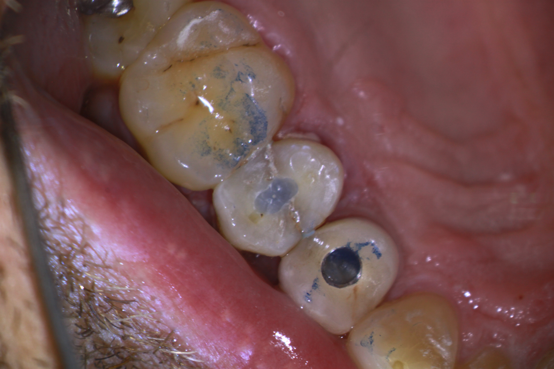
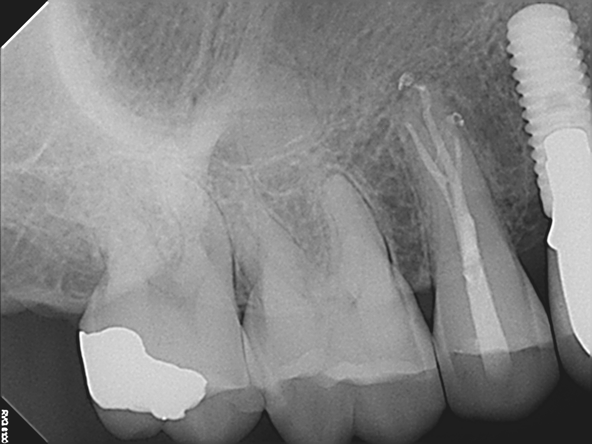
A 39 year old male patient arrived with a history of sharp chewing sensitivity and elevated thermal sensitivity in a maxillary 2nd premolar. He previously had the first premolar extracted and replaced with an implant. The referring Dentist could not clearly discern the root anatomy with conventional imaging.
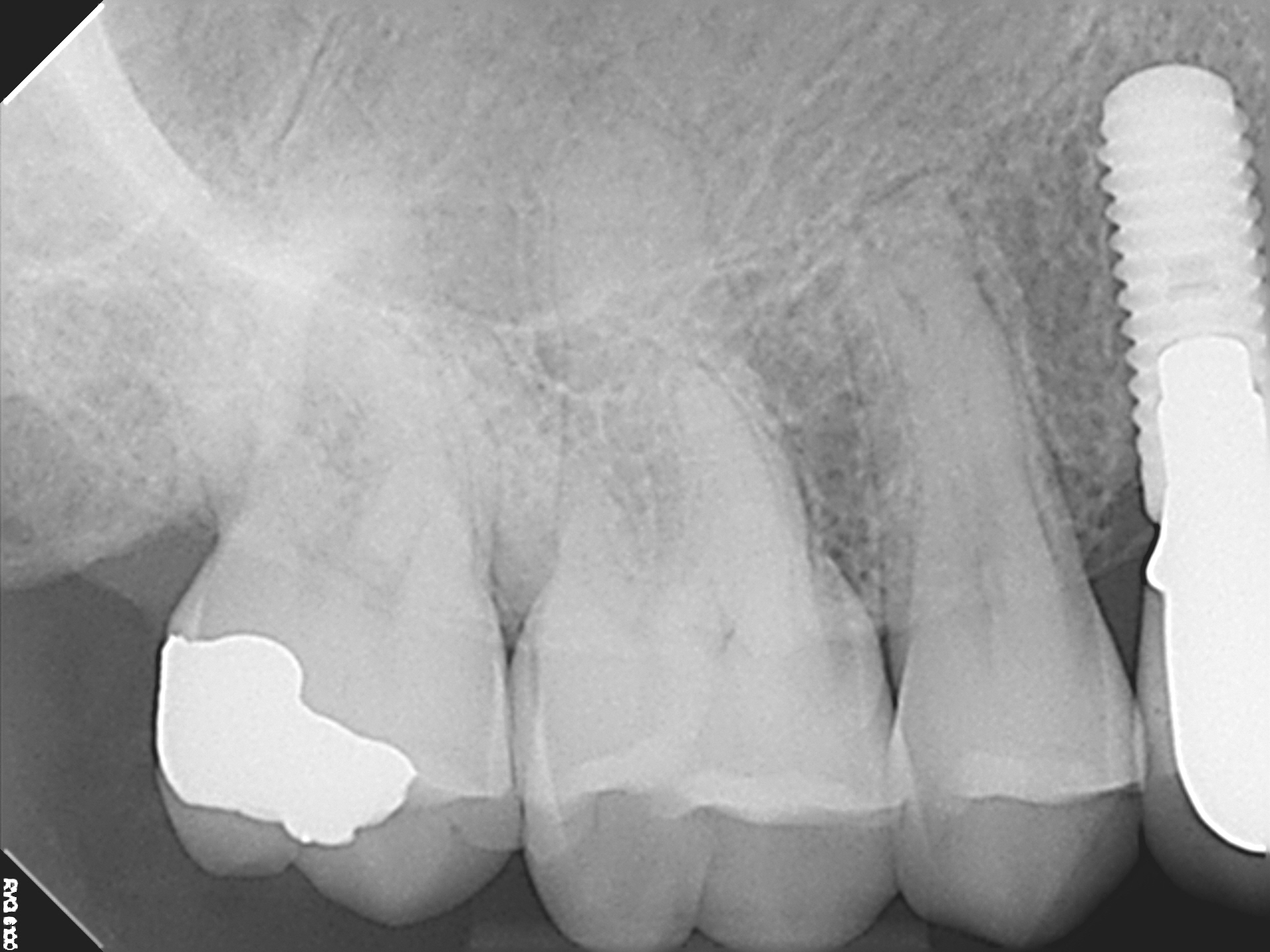
Preop image suggests multiple roots
Conventional imaging shows unusual canal anatomy that is unclear.
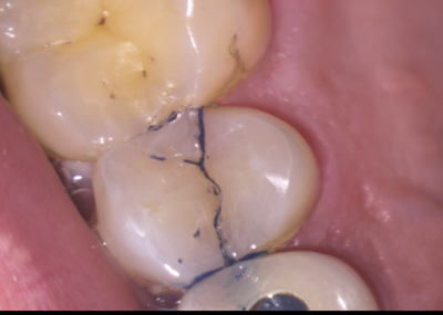
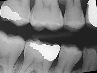
Virgin tooth shows mesial-distal crack that is easily stained with methylene blue dye. Tooth opposed by virgin premolar. Note adjacent premolar implant that MAY have been lost due to similar cracks. Is this a bruxism issue? Or perhaps an unlucky event with hard food?
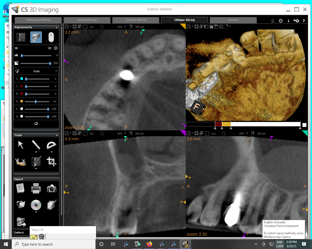
cbCT shows 3 canal premolar with 3 separate roots. Examination of the bite wing and adjacent proximal bone showed normal anatomy. This was consistent with the relatively normal proximal periodontal probings. This information not only allowed us to anticipate canal anatomy but it also helped to evaluate the prognosis. Although the tooth had cracked, the cracks did not appear severe enough at this stage for us to consider extraction rather than Endodontic treatment and restoration with full cuspal coverage,
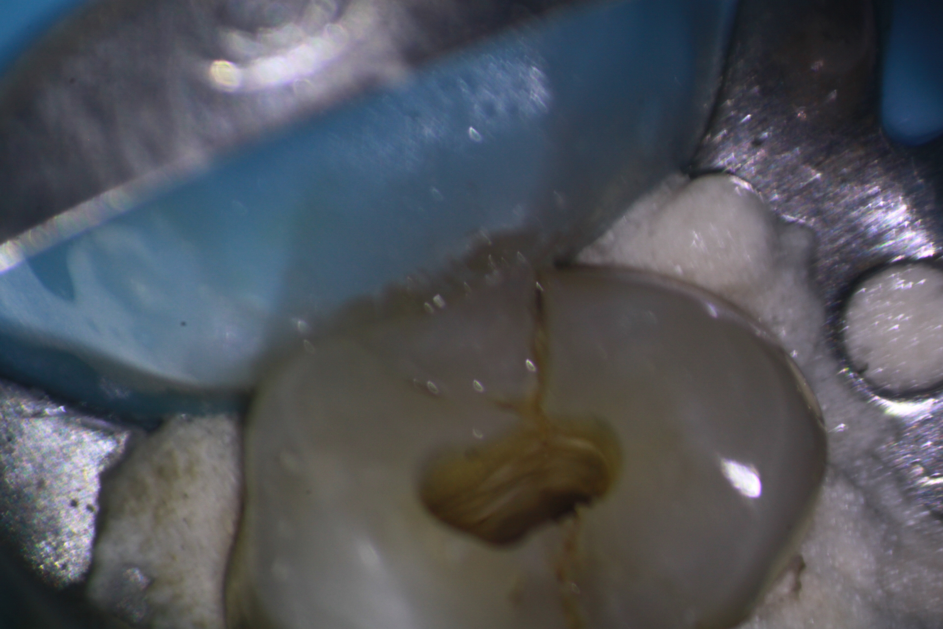
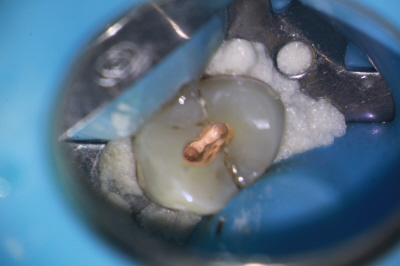
Off angle view of access showed the stained crack lines extending roughly to the CEJ. Final obturation with vertical compaction of warm gutta percha. Access sealed with Rebilda DC Bonded core paste. Patient advised to have crown placed STAT!!
