Adjacent Pathology Influences Treatment


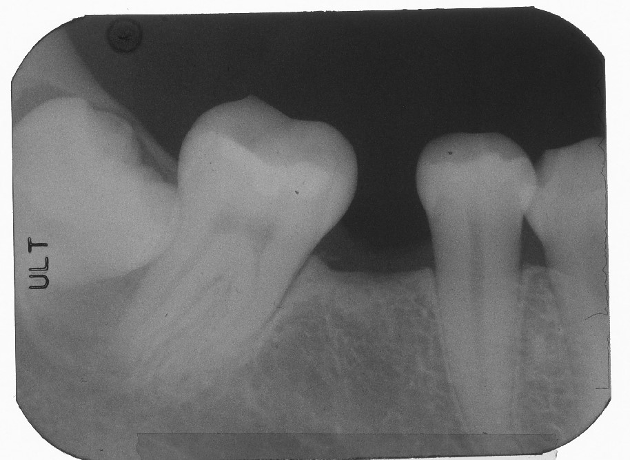
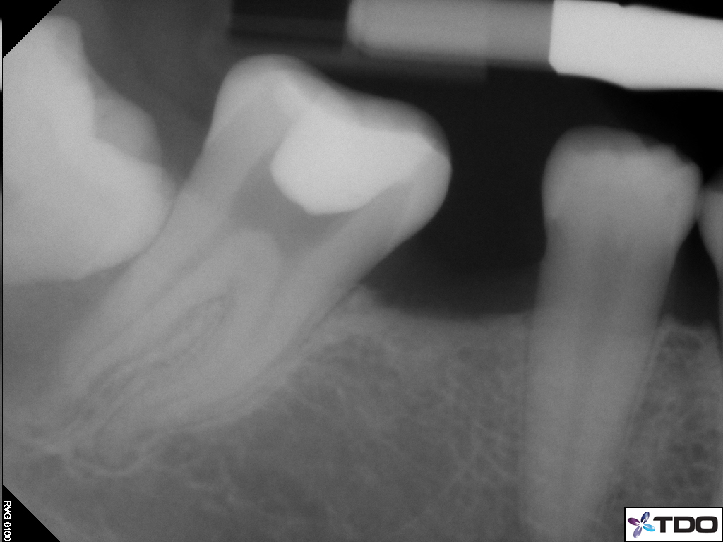
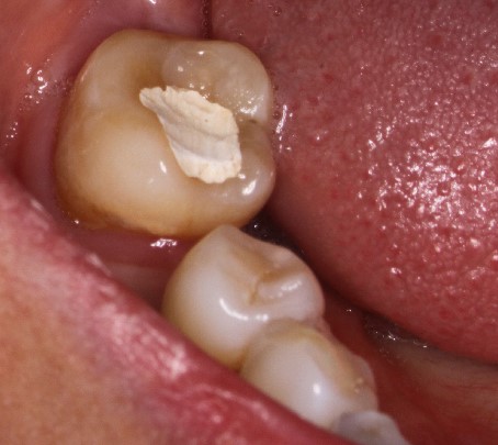
A 41 year old female patient was sent to me for continuation of treatment of tooth #47. The truth had been previously endodontically accessed by the referring Dentist become of symptoms in the area and a pulpectomy had been performed to get the patient comfortable. The referring dentist wished me to complete treatment. No other information was provided as to the original reason for treatment. Was a distal marginal ridge crack the reason for the endodontic intervention?
Clinical examination showed that was some percussion and chewing sensitivity in this tooth but this was consistent with the history of recently accessed incomplete endodontic treatment. However, the patient’s symptoms had not changed at all since the pulpectomy. Perio probings were normal. I was concerned that the problem may be related to the adjacent impacted wisdom tooth, #48, that also had a large radiolucent area associated with it as seen in the cbCT.
Prior to completion of Endodontic treatment of tooth #47, I recommended that tooth #48 be extracted. Once the extraction was complete symptoms needed to be re-evaluated. #47 also will need to be re-examined to determine whether there was enough post extraction distal bone support and what the long term Periodontal prognosis of #47 would be. Would it justify continuation of treatment?
The patient was referred to an Oral Surgeon to extract #48 as I had recommended. cbCT imaging was also sent and Endodontic treatment of #47 was delayed until further information was obtained from the Oral Surgeon.
The surgeon’s report included a surgical pathology report for the patient. It noted that the material removed from the pericoronal region around tooth #48 and running up the mid distal root surface of tooth #47 was consistent with a dentigerous cyst. Due to the size of the cyst, during removal, the oral surgeon found that there was no bone support for the distal surface of tooth #47 . Additionally, he was unable to remove the entirety of the cyst without removing the root of #47. For that reason BOTH teeth were removed during the surgery, as I originally suspected may be required. After the teeth were removed, the symptoms resolved and the area healed without incident. The oral surgeon was pleased with the results, given the size of the lesion and the proximity of the inferior alveolar nerve to the extraction site.
The patient will have an implant placed into position of tooth #46, which will be in better occlusal relationship with the opposing maxillary dentition.
This case teaches us a couple of things:
#1. The patient’s original symptom may not have been entirely related to problems with #47. Patients whose symptoms do not improve with initial emergency treatment can signal misdiagnosis.
We always need to take a more broad view of symptoms and consider whether associated or adjacent structures and pathology may be contributing to the patient’s current complaint.
#2. Where adjacent pathology (unknown radiolucency/Perio issues) threatens support for the tooth or treatment, it is prudent to deal with that pathology first because it frequently compromises the proposed endodontic treatment tooth. If the tooth is treated first, this may result in the previously Endo treated tooth being extracted and needless expense for the patient. Comprehensive treatment planning and assessment ensures that the right treatment is done, in the right sequence, the first time.
