Lateral Radiolucency
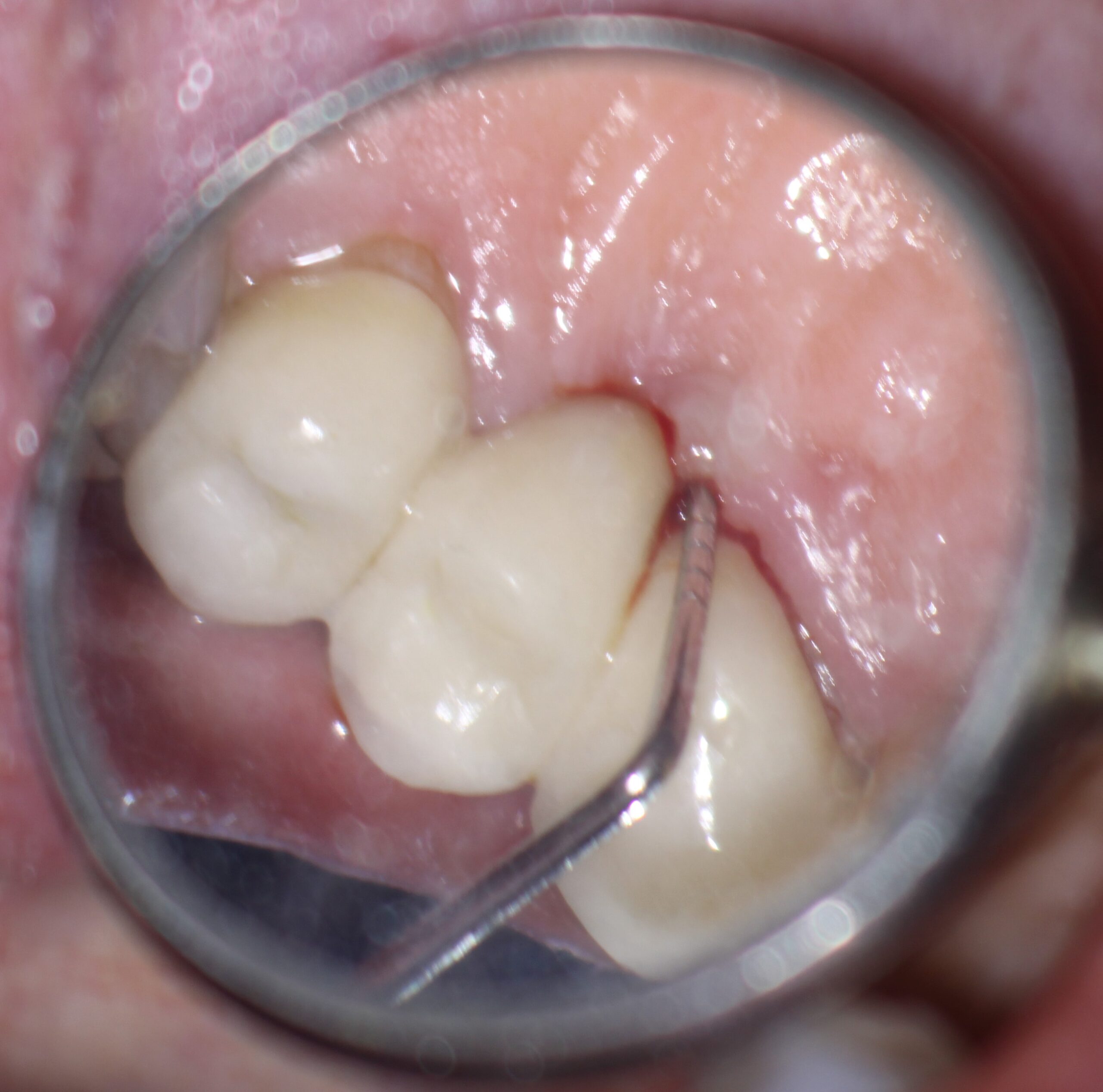
This 80 year old patient was sent to me with a history of draining buccal sinus associated with a maxillary right cuspid (#13). The tooth had been crowned as one of the abutments of a three unit bridge from #13 to #15. The patient was asymptomatic other than the draining sinus. Probings were within normal limits. Pulp tests showed no response in #13 .
Periapical radiography showed a large radiolucent finding associated with the apex of the cuspid as well as a large lateral radiolucent area on the mesial aspect that approximated the adjacent lateral incisor (#12). The radiolucency was quite large and although the lateral incisor appeared to be virgin, I wanted to make sure that the radiolucent finding was entirely associated with the cuspid and not with any pathology associated with the lateral incisor. Pulp tests of #12 showed normal responses .
The other consideration was Periodontal.Maybe this was a Perio lesion masquerading as Endo. Although there was a bit of bleeding on probing the palatal aspect of the cuspid, there were no deep pockets on the mesial or palatal side that suggested a cracked cuspid root or possible Perio issue causing the radiolucency.
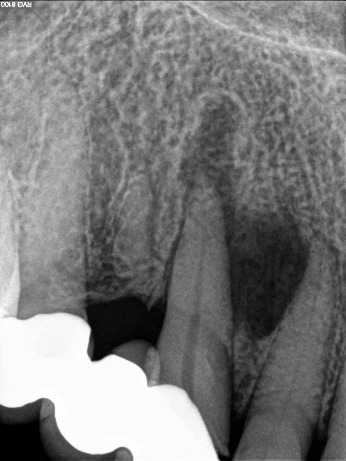
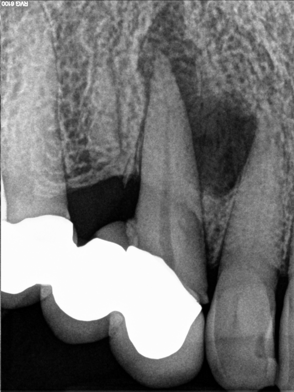
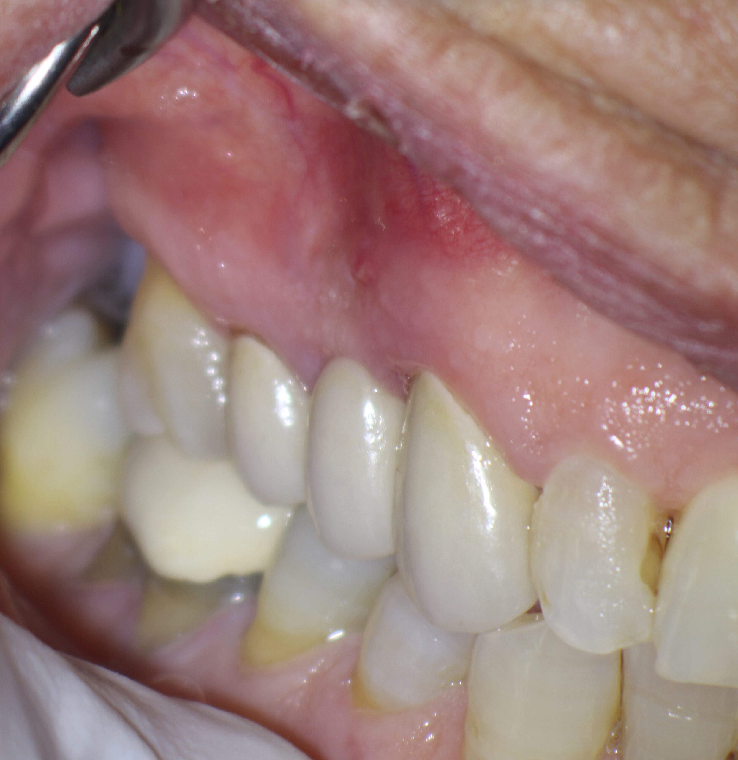
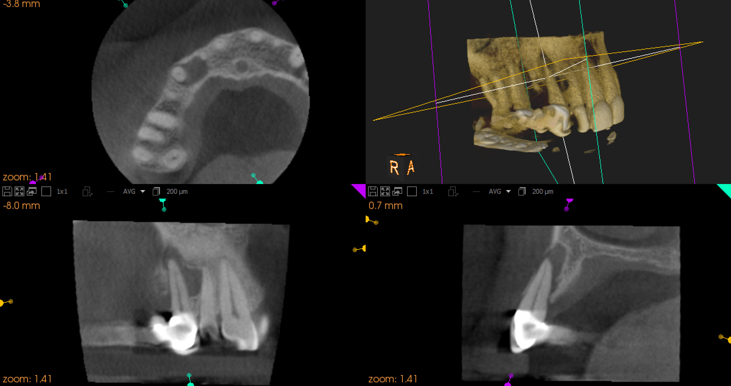
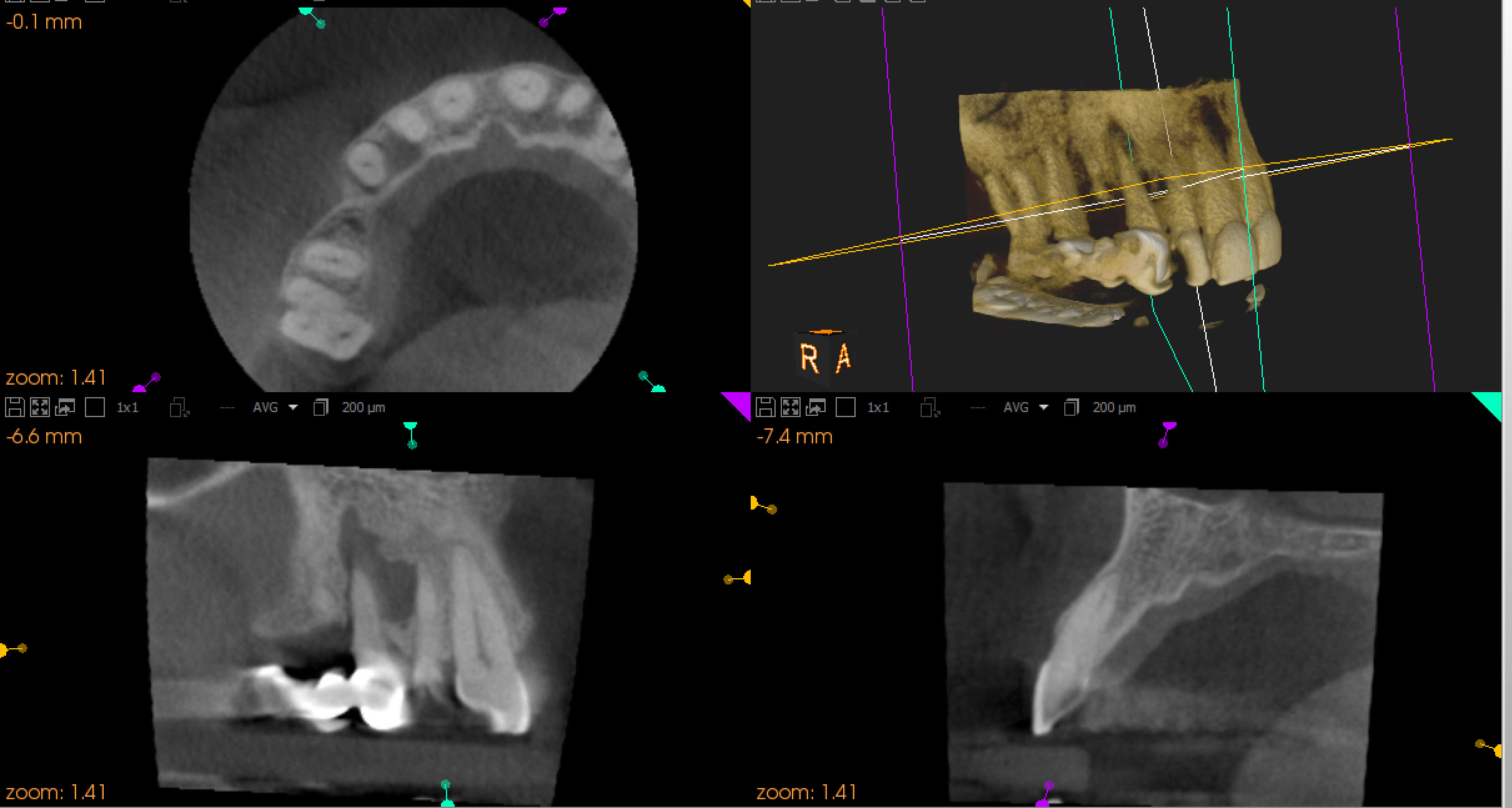
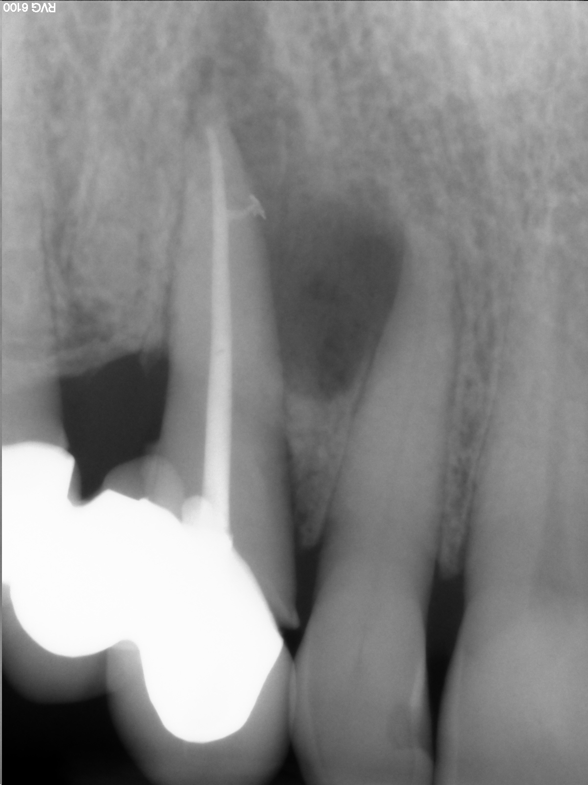
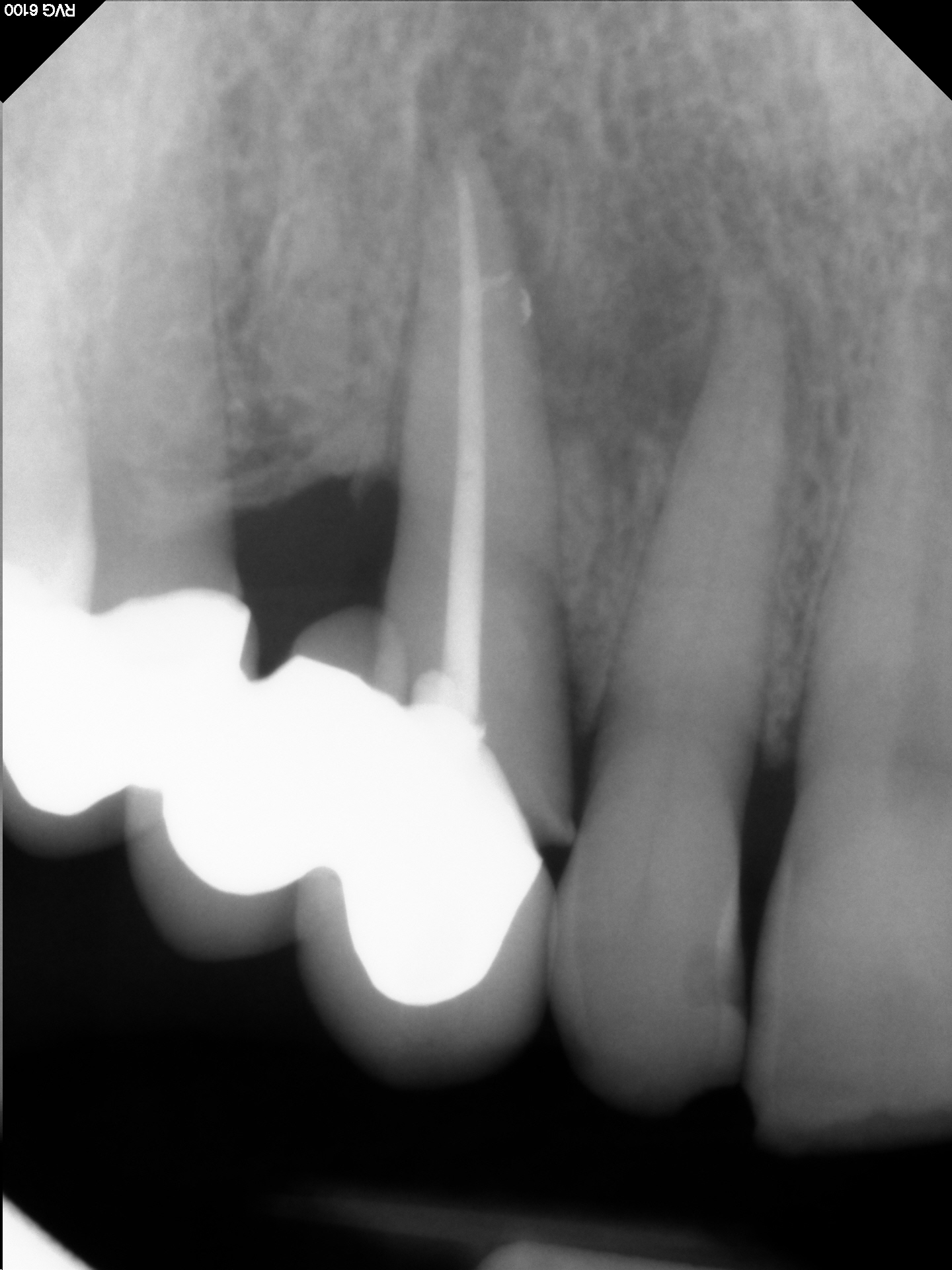
I explained to the patient that the pulp in the cuspid had gone non vital and that Endodontic treatment was required . I had some suspicions that there may be a lateral canal on the mesial aspect of the cuspid that was contributing to the large lateral radiolucency.
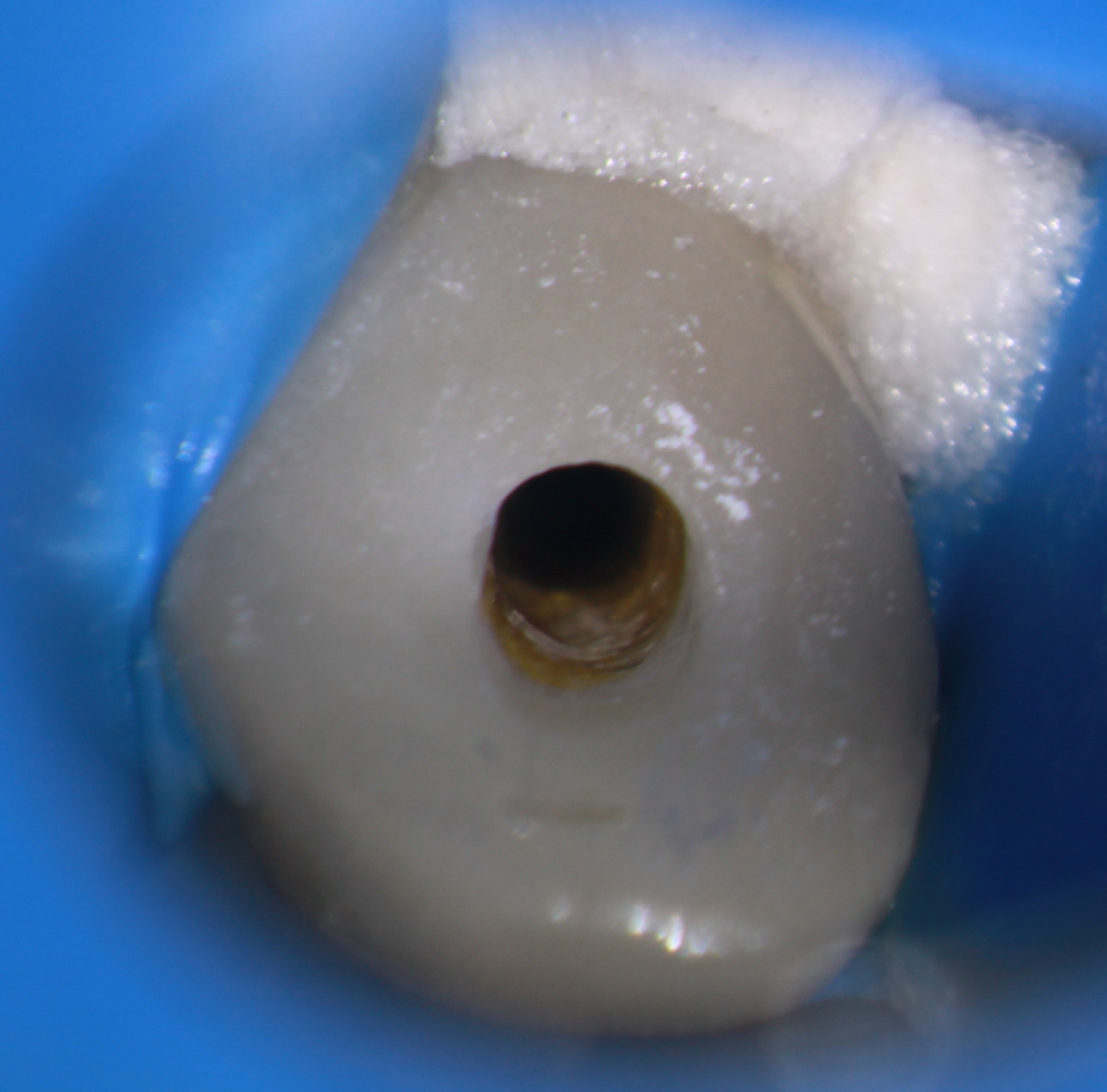
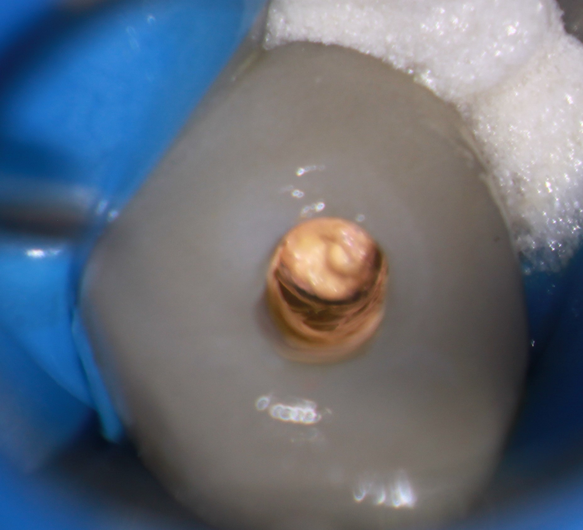
Access was made through the Crown and pulpal necrosis was confirmed. The canal was cleaned and shaped and the canal was medicated with calcium hydroxide. The patient was recalled a few weeks later at which time the draining sinus had closed and the tooth was ready for operation. The final operation results showed what we expected, a large filled lateral canal pointing directly to the lateral radiolucency on the mesial aspect of the root. Follow-up imaging at six months postop showed healing and regeneration of the bone associated with the cuspid.
What we can learn from this is that when the pulps of the adjacent teeth are determined to be normal and lateral radiolucencies occur in teeth with suspected necrotic pulps, we should expect some lateral anatomy that is ideally obturated with either gutta percha, sealer or a combination of the two . In any case adequate treatment of the canal system will result in healing of the attachment apparatus and resolution of the problem, as we see in this case .
